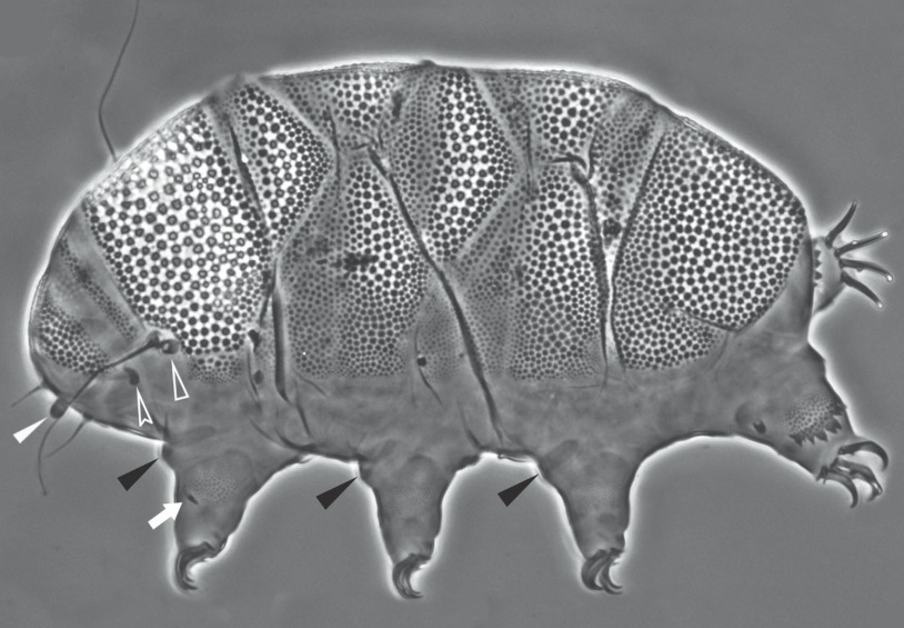
Acanthechiniscus

Acanthechiniscus, from Vecchi et al. 2016: “Echiniscid with pseudosegmental cuticular plate. Black eyes. Buccal cirri present, Filamentous cirri A present. Rigid buccal tube. Filaments and/or spines (may be small) present, generally in all lateral positions (B, C, D, E). Dorsal spines present. Notched collar (or spine fringe) on hind legs present.” Very similar to Pseudechiniscus, but notched cuff […]

Bryodelphax

From Ramazzotti & Maucci 1983 (tr. Beasley 1995): The median plates 1 and 2 are divided, not 3, so that there are visible total 5 median plates; lacking the notches on the terminal plate. From Kristensen 1987: “Sparsely pigmented Echiniscidae with red granulae eyes, unflexible buccal tube with CaCO3 encrusted stylet supports. Median plates 1 […]

Bryochoerus

From Ramazzotti & Maucci 1983 (tr. Beasley 1995): Median plates l, 2,3 divided transversely, so that total of 6 median plates visible. From Kristensen 1987: “Unpigmented to rose coloured Echiniscidae with red eyes, short inflexible buccal tube, with CaCO3 encrusted stylet supports. All median plates 1,2,3 divided, without pseudosegmental plates, with small lateral intersegmental plate […]

Testechiniscus

From Kristensen 1987: “Echiniscidae with black/brownish or ‘lipoid’ granular eyes; rigid buccal tube, large cuticular stylet supports, seven rows of sculptured ventral plates. Pseudosegmental plates absent. Paired segmental plates II and III with thinner anterior part. Caudal plate IV with a terminal indentation. Median plates 1 and 2 large, plate 3 small and reduced. Colour […]

Hypechiniscus

From Thulin 1928: “Echiniscidae with armor containing five median plates. End plate adjacent to the last median plate. Terminal notches present. Echiniscus gladiator MURRAY var. exarmatus MURR leads to this genus. The exarmatus form (from the Faroe Islands) that I have examined differs in some aspects from MURRAYS description: The animals are colorless with black […]

Echiniscus

From Schultze 1840: “The body ovate-elongated, with armor plates, in nine distinct segments, eight feet affixed to alternate segments from the third to the ninth. The head with four thorn-like antennae, eyes simple and two.” Untranslated: “Corpus ovato-elongatum, scutatum, spinosum, in novem segmenta distinctum, pedes octo alternis segmentis a tertio ad nonum affixi. Caput antennis […]

Diploechiniscus

From Vicente et al. 2013: “Echiniscids with dorsal plates I, II, III, IV (II and III paired), transversally subdivided median plates m1 and m2 and undivided plate m3 present; double sculpture in the dorsal plates, represented (under phase contrast) by dark polygonal and white circular grains; ventral plates present, especially evident in the anterior, head […]

Pseudechiniscus

Genus Pseudechiniscus From Ramazzotti & Maucci: “After the second paired plate, or after the third median plate if present, follows a pseudosegmental plate, paired or unpaired, and then the terminal plate; buccal cirri present; cirri A of filament shape.” From Kristensen 1987: “Echiniscidae with black eyes: rigid buccal canal, stylet supports may be present, but […]

Novechiniscus

From Kristensen 1987: “Echiniscidae with flexible buccal tube, pharyngeal bulb with a drop- shaped structure located on the placoids. Eyes possibly black, but nearly disappeared during the preparation. All four segmental plates I-IV unpaired with two bar-shaped median plates 1 and a distinct bar-shaped median plate 2; primary and secondary clavae with ring shaped annulus […]

Proechiniscus

Genus Proechiniscus From Kristensen 1987: “Echiniscidae with black eyes, flexible buccal canal, presence of calcified stylet supports. Segmental plates II and III paired with unpaired posterior element; median plates 1, 2 and 3 and the unpaired pseudosegmental plate IV’ bar- shaped; with dome-shaped secondary clavae. Colour reddish brown. Description: Proechinicus has unique dorsal plates. A series of […]

Claw Symmetry Relative To Median Plane Of Leg?

Image from Bingemer J, Hohberg K. 2017. An illustrated identification key to the eutardigrade species (Tardigrada, Eutardigrada) presently known from European soils. Soil Organisms. 89 (3): 127-149.

Stylet Support Insertion Point As Percentage Of Buccal Tube Length

Stylet support insertion point = ss divided by Buccal tube length, as %. Note anterior measurements begin at anterior margin of stylet sheaths, ss is centred where stylet supports reach buccal tube (this requires a good dorsal or lateral view for proper measurement)


Image from Tumanov DV. 2006. Five new species of the genus Milnesium (Tardigrada, Eutardigrada, Milnesiidae). Zootaxa. 1122: 1-23.

Buccal Tube Long, Mouth At Anterior Of A Protrusible Snout

Images from Pilato G, Binda MG. 2010. Definition of families, subfamilies, genera, and subgenera of the Eutardigrada, and keys to their identification. Zootaxa. 2404: 1-54.

Pharyngeal Tube With Spiral Reinforcement

Images from Pilato G, Binda MG. 2010. Definition of families, subfamilies, genera, and subgenera of the Eutardigrada, and keys to their identification. Zootaxa. 2404: 1-54.

Peribuccal papillae?

Note: in a tardigrade with snout extended, the flex of the pharyngeal tube may not be obvious.


Images from Pilato G, Binda MG. 2010. Definition of families, subfamilies, genera, and subgenera of the Eutardigrada, and keys to their identification. Zootaxa. 2404: 1-54.

(Lateral) cephalic papillae, peribuccal papillae, Milnesiidae - type claws, exceptionally wide (and usually short) buccal tube?

Cephalic image modified from Nelson DR, Guidetti R, Rebecchi L. 2009. Tardigrada. Ch. 14 in Ecology and Classification of North American Freshwater Invertebrates, Thorp JH, Covich AP (eds.), 3rd ed.

Claw image modified from Pilato G, Binda MG. 2010. Definition of families, subfamilies, genera, and subgenera of the Eutardigrada, and keys to their identification. Zootaxa. 2404: 1-54.

Pharyngeal tube flexible?

Note: in a tardigrade with snout extended, the flex of the pharyngeal tube may not be obvious.


Images from Pilato G, Binda MG. 2010. Definition of families, subfamilies, genera, and subgenera of the Eutardigrada, and keys to their identification. Zootaxa. 2404: 1-54.

Serrated / dentate cuff / collar on legs IV

Left image from Kristensen RM. 1987. Generic revision of the Echiniscidae (Heterotardigrada), with a discussion of the origin of the family. pp. 261-335 in Bertolani R (ed). Biology of Tardigrades: Selected symposia and monographs.

Right Image from Richters F. 1926. Tardigrada. in Krumbach T. 1927. Handbuch der Zoologie, 3rd band, Walter de Gruyter & Co.

Leg sensory structures

Typically papilla on leg IV (se4 in image) and variable shape on leg I


Image from Kristensen RM. 1987. Generic revision of the Echiniscidae (Heterotardigrada), with a discussion of the origin of the family. pp. 261-335 in Bertolani R (ed). Biology of Tardigrades: Selected symposia and monographs.

Trunk appendages

Appendages (after Cirrus A) labeled according to plate … B (Scapular plate), C (First segmental plate), D (Second segmental plate), E (terminal plate). “B” is lateral, “Bd” is dorsal

Be careful! There can be great variability within a population, with individuals (especially juveniles) lacking some appendages. When in doubt, choose “Unclear” to ignore this character!


Left image from Ramazzotti G, Maucci W. 1983. Il phylum Tardigrada(III edizione riveduta e aggiornata). English translation by C. W. Beasley, 1995. Memorie dell’ Istituto Italiano di Idrobiologia 41: 1-1012.

Right image cropped from Kristensen RM. 1987. Generic revision of the Echiniscidae (Heterotardigrada), with a discussion of the origin of the family. pp. 261-335 in Bertolani R (ed). Biology of Tardigrades: Selected symposia and monographs.

Cirrus A form

Images modified from Kristensen RM. 1987. Generic revision of the Echiniscidae (Heterotardigrada), with a discussion of the origin of the family. pp. 261-335 in Bertolani R (ed). Biology of Tardigrades: Selected symposia and monographs.

Lower image (Mopsechiniscus) modified from du Bois-Reymond Marcus E. 1944. Sobre tardigrados brasileiros. Communicaciones Zoologicas del Museo de Historia Natural de Montevideo. 1(13): 1-19 plus plates.

Terminal / caudal plate with two notches?

Images modified from Kristensen RM. 1987. Generic revision of the Echiniscidae (Heterotardigrada), with a discussion of the origin of the family. pp. 261-335 in Bertolani R (ed). Biology of Tardigrades: Selected symposia and monographs.

First image (Echiniscus) modified from Richters F. 1926. Tardigrada. in Krumbach T. 1927. Handbuch der Zoologie, 3rd band, Walter de Gruyter & Co.

Inner & outer buccal cirri present

Image modified from Nelson DR, Guidetti R, Rebecchi L. 2009. Tardigrada. Ch. 14 in Ecology and Classification of North American Freshwater Invertebrates, Thorp JH, Covich AP (eds.), 3rd ed.

Pseudosegmental plate between last medial plate & terminal plate?

Note on 2nd image, what you see before the terminal plate is either no plate, or a full-width medial plate; don’t confuse the latter with a pseudosegmental plate!
Also, sometimes the pseudosegmental plate is paired, like segmental plates II & III. It’s still pseudosegmental!

Images from Clifford HF. 1991. Aquatic invertebrates of Alberta. University of Alberta Press, Alberta, Canada.

Segmental plates larger than intersegmental?

Image modified from Lindahl & Balser (1999), http://www.iwu.edu/~tardisdp/Keypage35.html

Any Paired Plates?

Images from Kristensen RM. 1987. Generic revision of the Echiniscidae (Heterotardigrada), with a discussion of the origin of the family. pp. 261-335