Echiniscus

Richters F. 1926. Tardigrada. in Krumbach T. 1927. Handbuch der Zoologie, 3rd band, Walter de Gruyter & Co.

Class : Heterotardigrada

Order : Echiniscoidae

Family : Echiniscidae

Genus : Echiniscus

From Schultze 1840: “The body ovate-elongated, with armor plates, in nine distinct segments, eight feet affixed to alternate segments from the third to the ninth. The head with four thorn-like antennae, eyes simple and two.”

Untranslated: “Corpus ovato-elongatum, scutatum, spinosum, in novem segmenta distinctum, pedes octo alternis segmentis a tertio ad nonum affixi. Caput antennis quatuor spinisque duabus instructum, ocumli simplices duo.”

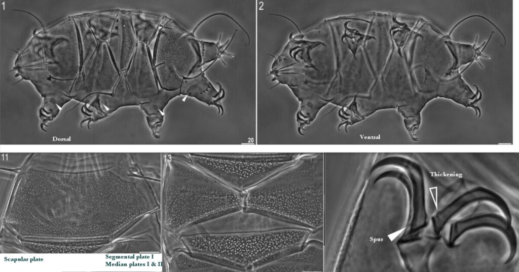
From Kristensen 1987: “Diagnosis: Echiniscidae with red-granulae eyes, unflexible buccal tube, without pseudosegmental plates IV’. Median plates undivided, caudal plate notched; ventral plates absent. Primary and secondary clavae papillate. Stylet supports absent or not visible by light microscopy. Emended description: The dorsal plates consist of a strongly sculptured head plate, indistinct neck plate, unpaired scapular plate (I), paired segmental plates II and III, unpaired notched caudal plate (IV), undivided plate 1 and 2; if present, me- dian plate 3 also undivided. Lateral intersegmental plates absent, segmental plates II and III may be divided into several pieces, as may the scapular plate (I). The leg plates are strongly developed only on the fourth pair of legs where the edge of the plate forms a serrated collar or a spine fringe. The cephalic sense organs consist of papillate primary and secondary clavae, long cirri A, short internal and slightly longer external cirri. The cirri A have small cirrophores separated from the primary clavae, cirrophores are present also on internal and external cirri. The leg I and 1V sense organs are present although they are not always illustrated in older descriptions. The leg I sense organ is located distally and it may vary from a short to a long spine; the leg IV sense organ is always in the form of a papilla located proximally on the leg. The arctomys and viridis groups lack the trunk appendages, but in other species, the location of spines or cirri have a high taxonomic value, and together with the sculpture of the dorsal plates, these constitute two of the best key characters in the determination of the species in the genus Echiniscus. The claw morphology is highly variable in the genus, accessory hooks, spurs or cusps may be absent as in the type species B. testudo, and the internal claws often have secondary spurs. In the more advanced species secondary and tertiary spurs can be present on the external claws, too. The location (proximal at the base of claw or more distal) also have a high taxonomic value. Colour varies widely e.g. grey, black, green, oliven, brown, red or yellow. The buccal apparatus has a relatively short, rigid buccal tube with stylets longer than the tube. The furcae of the stylets insert on the large, trilobed pharyngeal bulb. The stylet supports were not observed in any of the investigated species. The placoids and outer cuticular linings are well developed.”

From Gąsiorek et al. 2017: “Echiniscids with red eyes composed of pigment granules. Rigid buccal tube lacking stylet supports or with fine, fibrillar stylet supports. Two pairs of segmental plates. Two or three median plates, sometimes transversally subdivided. Incisions (notches) on caudal (terminal) plate. No pseudosegmental plates. Ventral subcephalic and/or genital plates may be present.

Differential diagnosis […phenotypic…] Echiniscus differs from the Pseudechiniscus– like genera (Acanthechiniscus Vecchi et al., 2016; Antechiniscus Kristensen, 1987; Cornechiniscus Maucci and Ramazzotti, 1981; Mopsechiniscus du Bois-Reymond Marcus, 1944; Multipseudechiniscus Schulte and Miller, 2011; Proechiniscus Kristensen, 1987; Pseudechiniscus Thulin, 1911) in having red eyes and lacking pseudosegmental plates (eyes are always crystalline and black, and at least one pseudosegmental plate occurs in these genera). The rigid buccal tube of Echiniscus differentiates the genus from Cornechiniscus, Mopsechiniscus, Multipseudechiniscus, Novechiniscus Kristensen, 1987, and Proechiniscus, in which the posterior portion of buccal tube is flexible (Miller et al., 2012). Moreover, Echiniscus differs from Novechiniscus and Parechiniscus Cuénot, 1926 in having paired segmental plates (whereas Novechiniscus and Parechiniscus have only single plates). Echiniscus can be separated from Bryodelphax Thulin, 1928 and Bryochoerus Marcus, 1936 by the presence of incisions on the caudal plate. Moreover, Bryodelphax and Bryochoerus are generally very small, with adults rarely exceeding 150 μm, whereas typical dimensions of Echiniscus adults are well above 200 μm. Echiniscus typically lacks ventral plates, which are present in Diploechiniscus Vicente et al., 2013 and Testechiniscus Kristensen, 1987. Finally, red granular eyes differentiate the genus from Diploechiniscus, Testechiniscus and Hypechiniscus Thulin, 1928, all of which have black crystalline eyes.”

Images modified from Gąsiorek P, Stec D, Morek W, Michalczyk Ł. 2017. An integrative redescription of Echiniscus testudo (Doyère, 1840), the nominal taxon for the class Heterotardigrada (Ecdysozoa: Panarthropoda: Tardigrada). Zoologischer Anzeiger. 270: 107-122.

Citations:

Gąsiorek P, Stec D, Morek W, Michalczyk Ł. 2017. An integrative redescription of Echiniscus testudo (Doyère, 1840), the nominal taxon for the class Heterotardigrada (Ecdysozoa: Panarthropoda: Tardigrada). Zoologischer Anzeiger. 270: 107-122.

Kristensen RM. 1987. Generic revision of the Echiniscidae (Heterotardigrada), with a discussion of the origin of the family. pp. 261-335 in Bertolani R (ed). Biology of Tardigrades: Selected symposia and monographs.

Schultze CAS. 1840. Echiniscus bellermanni, animal crustaceium, macrobioto hufelandi affine. Berolini, Berlin.

Claw Symmetry Relative To Median Plane Of Leg?

Image from Bingemer J, Hohberg K. 2017. An illustrated identification key to the eutardigrade species (Tardigrada, Eutardigrada) presently known from European soils. Soil Organisms. 89 (3): 127-149.

Stylet Support Insertion Point As Percentage Of Buccal Tube Length

Stylet support insertion point = ss divided by Buccal tube length, as %. Note anterior measurements begin at anterior margin of stylet sheaths, ss is centred where stylet supports reach buccal tube (this requires a good dorsal or lateral view for proper measurement)


Image from Tumanov DV. 2006. Five new species of the genus Milnesium (Tardigrada, Eutardigrada, Milnesiidae). Zootaxa. 1122: 1-23.

Buccal Tube Long, Mouth At Anterior Of A Protrusible Snout

Images from Pilato G, Binda MG. 2010. Definition of families, subfamilies, genera, and subgenera of the Eutardigrada, and keys to their identification. Zootaxa. 2404: 1-54.

Pharyngeal Tube With Spiral Reinforcement

Images from Pilato G, Binda MG. 2010. Definition of families, subfamilies, genera, and subgenera of the Eutardigrada, and keys to their identification. Zootaxa. 2404: 1-54.

Peribuccal papillae?

Note: in a tardigrade with snout extended, the flex of the pharyngeal tube may not be obvious.


Images from Pilato G, Binda MG. 2010. Definition of families, subfamilies, genera, and subgenera of the Eutardigrada, and keys to their identification. Zootaxa. 2404: 1-54.

(Lateral) cephalic papillae, peribuccal papillae, Milnesiidae - type claws, exceptionally wide (and usually short) buccal tube?

Cephalic image modified from Nelson DR, Guidetti R, Rebecchi L. 2009. Tardigrada. Ch. 14 in Ecology and Classification of North American Freshwater Invertebrates, Thorp JH, Covich AP (eds.), 3rd ed.

Claw image modified from Pilato G, Binda MG. 2010. Definition of families, subfamilies, genera, and subgenera of the Eutardigrada, and keys to their identification. Zootaxa. 2404: 1-54.

Pharyngeal tube flexible?

Note: in a tardigrade with snout extended, the flex of the pharyngeal tube may not be obvious.


Images from Pilato G, Binda MG. 2010. Definition of families, subfamilies, genera, and subgenera of the Eutardigrada, and keys to their identification. Zootaxa. 2404: 1-54.

Serrated / dentate cuff / collar on legs IV

Left image from Kristensen RM. 1987. Generic revision of the Echiniscidae (Heterotardigrada), with a discussion of the origin of the family. pp. 261-335 in Bertolani R (ed). Biology of Tardigrades: Selected symposia and monographs.

Right Image from Richters F. 1926. Tardigrada. in Krumbach T. 1927. Handbuch der Zoologie, 3rd band, Walter de Gruyter & Co.

Leg sensory structures

Typically papilla on leg IV (se4 in image) and variable shape on leg I


Image from Kristensen RM. 1987. Generic revision of the Echiniscidae (Heterotardigrada), with a discussion of the origin of the family. pp. 261-335 in Bertolani R (ed). Biology of Tardigrades: Selected symposia and monographs.

Trunk appendages

Appendages (after Cirrus A) labeled according to plate … B (Scapular plate), C (First segmental plate), D (Second segmental plate), E (terminal plate). “B” is lateral, “Bd” is dorsal

Be careful! There can be great variability within a population, with individuals (especially juveniles) lacking some appendages. When in doubt, choose “Unclear” to ignore this character!


Left image from Ramazzotti G, Maucci W. 1983. Il phylum Tardigrada(III edizione riveduta e aggiornata). English translation by C. W. Beasley, 1995. Memorie dell’ Istituto Italiano di Idrobiologia 41: 1-1012.

Right image cropped from Kristensen RM. 1987. Generic revision of the Echiniscidae (Heterotardigrada), with a discussion of the origin of the family. pp. 261-335 in Bertolani R (ed). Biology of Tardigrades: Selected symposia and monographs.

Cirrus A form

Images modified from Kristensen RM. 1987. Generic revision of the Echiniscidae (Heterotardigrada), with a discussion of the origin of the family. pp. 261-335 in Bertolani R (ed). Biology of Tardigrades: Selected symposia and monographs.

Lower image (Mopsechiniscus) modified from du Bois-Reymond Marcus E. 1944. Sobre tardigrados brasileiros. Communicaciones Zoologicas del Museo de Historia Natural de Montevideo. 1(13): 1-19 plus plates.

Terminal / caudal plate with two notches?

Images modified from Kristensen RM. 1987. Generic revision of the Echiniscidae (Heterotardigrada), with a discussion of the origin of the family. pp. 261-335 in Bertolani R (ed). Biology of Tardigrades: Selected symposia and monographs.

First image (Echiniscus) modified from Richters F. 1926. Tardigrada. in Krumbach T. 1927. Handbuch der Zoologie, 3rd band, Walter de Gruyter & Co.

Inner & outer buccal cirri present

Image modified from Nelson DR, Guidetti R, Rebecchi L. 2009. Tardigrada. Ch. 14 in Ecology and Classification of North American Freshwater Invertebrates, Thorp JH, Covich AP (eds.), 3rd ed.

Pseudosegmental plate between last medial plate & terminal plate?

Note on 2nd image, what you see before the terminal plate is either no plate, or a full-width medial plate; don’t confuse the latter with a pseudosegmental plate!
Also, sometimes the pseudosegmental plate is paired, like segmental plates II & III. It’s still pseudosegmental!

Images from Clifford HF. 1991. Aquatic invertebrates of Alberta. University of Alberta Press, Alberta, Canada.

Segmental plates larger than intersegmental?

Image modified from Lindahl & Balser (1999), http://www.iwu.edu/~tardisdp/Keypage35.html

Any Paired Plates?

Images from Kristensen RM. 1987. Generic revision of the Echiniscidae (Heterotardigrada), with a discussion of the origin of the family. pp. 261-335