Diploechiniscus

Vicente F, Fontoura P, Cesari M, Rebecchi L, Guidetti R, Serrano A, Bertolani R. 2013. Integrative taxonomy allows the identification of synonymous species and the erection of a new genus of Echiniscidae (Tardigrada, Heterotardigrada). Zootaxa. 361396): 557-572.

Class : Heterotardigrada

Order : Echiniscoidae

Family : Echiiniscidae

Genus : Diploechiiniscus

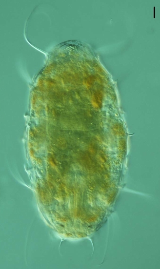
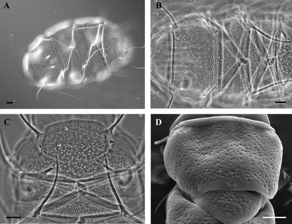
From Vicente et al. 2013: “Echiniscids with dorsal plates I, II, III, IV (II and III paired), transversally subdivided median plates m1 and m2 and undivided plate m3 present; double sculpture in the dorsal plates, represented (under phase contrast) by dark polygonal and white circular grains; ventral plates present, especially evident in the anterior, head region and around the gonopore; supernumerary dorsal-lateral spines present; buccal tube long and narrow, with stylet supports. Orange body, dark-brown eyes.”

ibid: “Re-description of the species (from the original description and from re-examined specimens collected in Forså, Norway; Sierra de Urbion, Spain; Caldas das Taipas, Vilar Formoso, Castro Laboreiro and Moita do Conqueiro, Portugal). Body colour orange. Eye spots simple and dark brown. Buccal cirri long, clavae large. Stylet supports present (sometimes difficult to observe in older slides). Dorsal plates present, all (except neck plate) characterized by double sculpture, which appears as dark, regular polygonal grains under white circular grains when viewed with phase contrast. Dark grains are separated by thin, white region from neighbours (normally groups of six); white grains of various sizes, never overlaping dark grains, and irregularly distributed. Cephalic plate unpaired, with median depression to the anterior margin; fine anterior sculpture and larger posterior double sculpture. Neck plate, a long transverse and relatively thin band, anterior and posterior region unsculptured and fine, dark grains in the middle. Dorsal segmental plates: plate I (or scapular plate) entire, with two sculptured small lateral plates exhibiting fine, dark grains; plates II and III paired and characterised by an unsculptured transverse band, and plate IV (or terminal plate), entire but faceted and notched (not obvious in older specimens). Median intersegmental plates: plate m1, transversally subdivided, anterior region formed of a large, flat and thin rectangle not always obvious due to overlapping scapular plate; plate m2, transversally subdivided and with an unsculptured transverse band, plate appears as two obtuse angle isosceles triangles joined by their larger side; and m3, entire, small and not obvious but with double sculpture. Lateral intersegmental plates are difficult to identify, though unsculptured spaces exist at la2 and la3. Long filaments A, B, C, D and E, sometimes barbed. Short hooked dorsal-lateral spines B’, C’, D’ and E’. Lateral spine E’, simple or double. Bd variable as long spine, very short spur, or absent and can be present on one or both sides of plate II. Long filaments Cd and short spines Dd. Ventral sculpture present as fine granulation, with clearly visible head plate and posterior plates beside gonopore. Leg plates present laterally, with dark granular sculpture. Spiniform papilla present on leg I; papilla on leg IV with rounded tip. Hooked spurs on all internal claws, external claws I–III smooth, occasionally one or two short right-angled spurs on the leg IV. Dentate collar variable, comprised of six to 13 triangular teeth, some irregularly bifurcated. Gonopore; a short tube in the males, and rosette in the females. The geographical distribution of E. oihonnae includes: Portugal, Switzerland, Northern Europe (including polar islands), U.S.A., Canada, Australia (Ramazzotti & Maucci 1983); Japan (Mathews, 1937); Kuril Islands, Far East Russia (Dudichev & Biserov 2000). Most of the non-European citations require confirmation, as for example, Murray (1910) was doubtful about his identification of Australian and Canadian specimens, and the Californian specimens, initially assigned to E. oihonnae, were revised as T. laterculus (Schuster, Grigarick & Toftner, 1980).”

Vicente F, Fontoura P, Cesari M, Rebecchi L, Guidetti R, Serrano A, Bertolani R. 2013. Integrative taxonomy allows the identification of synonymous species and the erection of a new genus of Echiniscidae (Tardigrada, Heterotardigrada). Zootaxa. 361396): 557-572.

Citations:

Vicente F, Fontoura P, Cesari M, Rebecchi L, Guidetti R, Serrano A, Bertolani R. 2013. Integrative taxonomy allows the identification of synonymous species and the erection of a new genus of Echiniscidae (Tardigrada, Heterotardigrada). Zootaxa. 361396): 557-572.

Claw Symmetry Relative To Median Plane Of Leg?

Image from Bingemer J, Hohberg K. 2017. An illustrated identification key to the eutardigrade species (Tardigrada, Eutardigrada) presently known from European soils. Soil Organisms. 89 (3): 127-149.

Stylet Support Insertion Point As Percentage Of Buccal Tube Length

Stylet support insertion point = ss divided by Buccal tube length, as %. Note anterior measurements begin at anterior margin of stylet sheaths, ss is centred where stylet supports reach buccal tube (this requires a good dorsal or lateral view for proper measurement)


Image from Tumanov DV. 2006. Five new species of the genus Milnesium (Tardigrada, Eutardigrada, Milnesiidae). Zootaxa. 1122: 1-23.

Buccal Tube Long, Mouth At Anterior Of A Protrusible Snout

Images from Pilato G, Binda MG. 2010. Definition of families, subfamilies, genera, and subgenera of the Eutardigrada, and keys to their identification. Zootaxa. 2404: 1-54.

Pharyngeal Tube With Spiral Reinforcement

Images from Pilato G, Binda MG. 2010. Definition of families, subfamilies, genera, and subgenera of the Eutardigrada, and keys to their identification. Zootaxa. 2404: 1-54.

Peribuccal papillae?

Note: in a tardigrade with snout extended, the flex of the pharyngeal tube may not be obvious.


Images from Pilato G, Binda MG. 2010. Definition of families, subfamilies, genera, and subgenera of the Eutardigrada, and keys to their identification. Zootaxa. 2404: 1-54.

(Lateral) cephalic papillae, peribuccal papillae, Milnesiidae - type claws, exceptionally wide (and usually short) buccal tube?

Cephalic image modified from Nelson DR, Guidetti R, Rebecchi L. 2009. Tardigrada. Ch. 14 in Ecology and Classification of North American Freshwater Invertebrates, Thorp JH, Covich AP (eds.), 3rd ed.

Claw image modified from Pilato G, Binda MG. 2010. Definition of families, subfamilies, genera, and subgenera of the Eutardigrada, and keys to their identification. Zootaxa. 2404: 1-54.

Pharyngeal tube flexible?

Note: in a tardigrade with snout extended, the flex of the pharyngeal tube may not be obvious.


Images from Pilato G, Binda MG. 2010. Definition of families, subfamilies, genera, and subgenera of the Eutardigrada, and keys to their identification. Zootaxa. 2404: 1-54.

Serrated / dentate cuff / collar on legs IV

Left image from Kristensen RM. 1987. Generic revision of the Echiniscidae (Heterotardigrada), with a discussion of the origin of the family. pp. 261-335 in Bertolani R (ed). Biology of Tardigrades: Selected symposia and monographs.

Right Image from Richters F. 1926. Tardigrada. in Krumbach T. 1927. Handbuch der Zoologie, 3rd band, Walter de Gruyter & Co.

Leg sensory structures

Typically papilla on leg IV (se4 in image) and variable shape on leg I


Image from Kristensen RM. 1987. Generic revision of the Echiniscidae (Heterotardigrada), with a discussion of the origin of the family. pp. 261-335 in Bertolani R (ed). Biology of Tardigrades: Selected symposia and monographs.

Trunk appendages

Appendages (after Cirrus A) labeled according to plate … B (Scapular plate), C (First segmental plate), D (Second segmental plate), E (terminal plate). “B” is lateral, “Bd” is dorsal

Be careful! There can be great variability within a population, with individuals (especially juveniles) lacking some appendages. When in doubt, choose “Unclear” to ignore this character!


Left image from Ramazzotti G, Maucci W. 1983. Il phylum Tardigrada(III edizione riveduta e aggiornata). English translation by C. W. Beasley, 1995. Memorie dell’ Istituto Italiano di Idrobiologia 41: 1-1012.

Right image cropped from Kristensen RM. 1987. Generic revision of the Echiniscidae (Heterotardigrada), with a discussion of the origin of the family. pp. 261-335 in Bertolani R (ed). Biology of Tardigrades: Selected symposia and monographs.

Cirrus A form

Images modified from Kristensen RM. 1987. Generic revision of the Echiniscidae (Heterotardigrada), with a discussion of the origin of the family. pp. 261-335 in Bertolani R (ed). Biology of Tardigrades: Selected symposia and monographs.

Lower image (Mopsechiniscus) modified from du Bois-Reymond Marcus E. 1944. Sobre tardigrados brasileiros. Communicaciones Zoologicas del Museo de Historia Natural de Montevideo. 1(13): 1-19 plus plates.

Terminal / caudal plate with two notches?

Images modified from Kristensen RM. 1987. Generic revision of the Echiniscidae (Heterotardigrada), with a discussion of the origin of the family. pp. 261-335 in Bertolani R (ed). Biology of Tardigrades: Selected symposia and monographs.

First image (Echiniscus) modified from Richters F. 1926. Tardigrada. in Krumbach T. 1927. Handbuch der Zoologie, 3rd band, Walter de Gruyter & Co.

Inner & outer buccal cirri present

Image modified from Nelson DR, Guidetti R, Rebecchi L. 2009. Tardigrada. Ch. 14 in Ecology and Classification of North American Freshwater Invertebrates, Thorp JH, Covich AP (eds.), 3rd ed.

Pseudosegmental plate between last medial plate & terminal plate?

Note on 2nd image, what you see before the terminal plate is either no plate, or a full-width medial plate; don’t confuse the latter with a pseudosegmental plate!
Also, sometimes the pseudosegmental plate is paired, like segmental plates II & III. It’s still pseudosegmental!

Images from Clifford HF. 1991. Aquatic invertebrates of Alberta. University of Alberta Press, Alberta, Canada.

Segmental plates larger than intersegmental?

Image modified from Lindahl & Balser (1999), http://www.iwu.edu/~tardisdp/Keypage35.html

Any Paired Plates?

Images from Kristensen RM. 1987. Generic revision of the Echiniscidae (Heterotardigrada), with a discussion of the origin of the family. pp. 261-335