Bryodelphax

du Bois-Reymond Marcus E. 1944. Sobre tardigrados brasileiros. Communicaciones Zoologicas del Museo de Historia Natural de Montevideo. 1(13): 1-19 plus plates.

Class : Heterotardigrada

Order : Echiniscoidae

Family : Echiniscidae

Genus : Bryodelphax

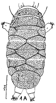
From Ramazzotti & Maucci 1983 (tr. Beasley 1995): The median plates 1 and 2 are divided, not 3, so that there are visible total 5 median plates; lacking the notches on the terminal plate.

From Kristensen 1987: “Sparsely pigmented Echiniscidae with red granulae eyes, unflexible buccal tube with CaCO3 encrusted stylet supports. Median plates 1 and 2 divid- ed. Median plate 3 undivided with round to straight posterior margin. Without any form for pseudosegmentation. Ventral plates can be present.

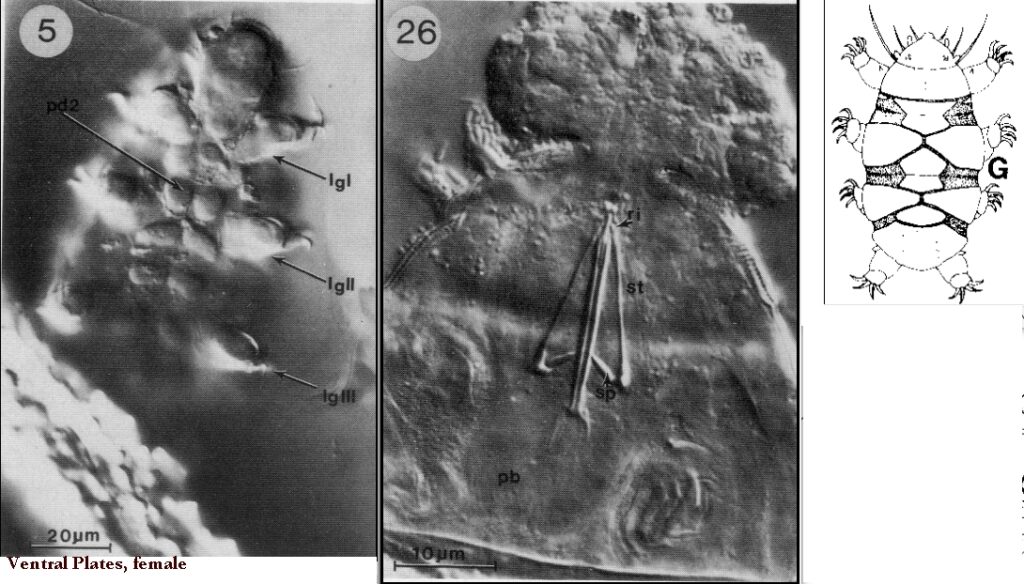
Emended description: Ventral plates present in at least two species (B. sinensis and B. weglarskae). Moreover, indistinct ventral plates, especially two small genital plates, may be present in B. parvulas. (Note: the presence of the thin ventral plates can be overlooked in some species of Bryodelphax, if the prepara- tion is not polyvinyl—lactophenol). The dorsal plate has a finely punctuated cuti- cle (pillars present in the epicuticle) and light round area (depression in the cuticle). Dark granulation (elevations in the cuticle) is seen in a few species. The head and neck plates are indistinct. The scapular plate (I) is large, paired segmental plates II and III are often divided into an anterior and posterior part. Caudal plate (IV) without terminal indentation (notches), but two lateral eleva- tions or facets (pseudonotches) in the cuticle are present. Median plates 1 and 2 are divided into two parts, the median plate 3 is undivided, its posterior margin of median plate 3 is often beneath the caudal plate. The cephalic appendages consist of the Echiniscus-type with both primary and secondary clavae formed as straight, small papillae. The cirri are short and cirrophores were not seen. The male, which is the same size at the female, does not have enlarged clavae. Trunk cirri are absent. Sense organs present on legs I and IV. Leg plate may be absent on all legs, often present as a serrated collar only on leg IV. A character unique for the genus Bryodelphax is 10 small buccal papillae around the mouth opening. The buccal apparatus and claws are similar to those of Bryochoerus.”

Images cropped from Kristensen RM. 1987. Generic revision of the Echiniscidae (Heterotardigrada), with a discussion of the origin of the family. pp. 261-335 in Bertolani R (ed). Biology of Tardigrades: Selected symposia and monographs.

Citations:

Kristensen RM. 1987. Generic revision of the Echiniscidae (Heterotardigrada), with a discussion of the origin of the family. pp. 261-335 in Bertolani R (ed). Biology of Tardigrades: Selected symposia and monographs.

Ramazzotti G, Maucci W. 1983. Il phylum Tardigrada(III edizione riveduta e aggiornata). English translation by C. W. Beasley, 1995. Memorie dell’ Istituto Italiano di Idrobiologia 41: 1-1012.

Claw Symmetry Relative To Median Plane Of Leg?

Image from Bingemer J, Hohberg K. 2017. An illustrated identification key to the eutardigrade species (Tardigrada, Eutardigrada) presently known from European soils. Soil Organisms. 89 (3): 127-149.

Stylet Support Insertion Point As Percentage Of Buccal Tube Length

Stylet support insertion point = ss divided by Buccal tube length, as %. Note anterior measurements begin at anterior margin of stylet sheaths, ss is centred where stylet supports reach buccal tube (this requires a good dorsal or lateral view for proper measurement)


Image from Tumanov DV. 2006. Five new species of the genus Milnesium (Tardigrada, Eutardigrada, Milnesiidae). Zootaxa. 1122: 1-23.

Buccal Tube Long, Mouth At Anterior Of A Protrusible Snout

Images from Pilato G, Binda MG. 2010. Definition of families, subfamilies, genera, and subgenera of the Eutardigrada, and keys to their identification. Zootaxa. 2404: 1-54.

Pharyngeal Tube With Spiral Reinforcement

Images from Pilato G, Binda MG. 2010. Definition of families, subfamilies, genera, and subgenera of the Eutardigrada, and keys to their identification. Zootaxa. 2404: 1-54.

Peribuccal papillae?

Note: in a tardigrade with snout extended, the flex of the pharyngeal tube may not be obvious.


Images from Pilato G, Binda MG. 2010. Definition of families, subfamilies, genera, and subgenera of the Eutardigrada, and keys to their identification. Zootaxa. 2404: 1-54.

(Lateral) cephalic papillae, peribuccal papillae, Milnesiidae - type claws, exceptionally wide (and usually short) buccal tube?

Cephalic image modified from Nelson DR, Guidetti R, Rebecchi L. 2009. Tardigrada. Ch. 14 in Ecology and Classification of North American Freshwater Invertebrates, Thorp JH, Covich AP (eds.), 3rd ed.

Claw image modified from Pilato G, Binda MG. 2010. Definition of families, subfamilies, genera, and subgenera of the Eutardigrada, and keys to their identification. Zootaxa. 2404: 1-54.

Pharyngeal tube flexible?

Note: in a tardigrade with snout extended, the flex of the pharyngeal tube may not be obvious.


Images from Pilato G, Binda MG. 2010. Definition of families, subfamilies, genera, and subgenera of the Eutardigrada, and keys to their identification. Zootaxa. 2404: 1-54.

Serrated / dentate cuff / collar on legs IV

Left image from Kristensen RM. 1987. Generic revision of the Echiniscidae (Heterotardigrada), with a discussion of the origin of the family. pp. 261-335 in Bertolani R (ed). Biology of Tardigrades: Selected symposia and monographs.

Right Image from Richters F. 1926. Tardigrada. in Krumbach T. 1927. Handbuch der Zoologie, 3rd band, Walter de Gruyter & Co.

Leg sensory structures

Typically papilla on leg IV (se4 in image) and variable shape on leg I


Image from Kristensen RM. 1987. Generic revision of the Echiniscidae (Heterotardigrada), with a discussion of the origin of the family. pp. 261-335 in Bertolani R (ed). Biology of Tardigrades: Selected symposia and monographs.

Trunk appendages

Appendages (after Cirrus A) labeled according to plate … B (Scapular plate), C (First segmental plate), D (Second segmental plate), E (terminal plate). “B” is lateral, “Bd” is dorsal

Be careful! There can be great variability within a population, with individuals (especially juveniles) lacking some appendages. When in doubt, choose “Unclear” to ignore this character!


Left image from Ramazzotti G, Maucci W. 1983. Il phylum Tardigrada(III edizione riveduta e aggiornata). English translation by C. W. Beasley, 1995. Memorie dell’ Istituto Italiano di Idrobiologia 41: 1-1012.

Right image cropped from Kristensen RM. 1987. Generic revision of the Echiniscidae (Heterotardigrada), with a discussion of the origin of the family. pp. 261-335 in Bertolani R (ed). Biology of Tardigrades: Selected symposia and monographs.

Cirrus A form

Images modified from Kristensen RM. 1987. Generic revision of the Echiniscidae (Heterotardigrada), with a discussion of the origin of the family. pp. 261-335 in Bertolani R (ed). Biology of Tardigrades: Selected symposia and monographs.

Lower image (Mopsechiniscus) modified from du Bois-Reymond Marcus E. 1944. Sobre tardigrados brasileiros. Communicaciones Zoologicas del Museo de Historia Natural de Montevideo. 1(13): 1-19 plus plates.

Terminal / caudal plate with two notches?

Images modified from Kristensen RM. 1987. Generic revision of the Echiniscidae (Heterotardigrada), with a discussion of the origin of the family. pp. 261-335 in Bertolani R (ed). Biology of Tardigrades: Selected symposia and monographs.

First image (Echiniscus) modified from Richters F. 1926. Tardigrada. in Krumbach T. 1927. Handbuch der Zoologie, 3rd band, Walter de Gruyter & Co.

Inner & outer buccal cirri present

Image modified from Nelson DR, Guidetti R, Rebecchi L. 2009. Tardigrada. Ch. 14 in Ecology and Classification of North American Freshwater Invertebrates, Thorp JH, Covich AP (eds.), 3rd ed.

Pseudosegmental plate between last medial plate & terminal plate?

Note on 2nd image, what you see before the terminal plate is either no plate, or a full-width medial plate; don’t confuse the latter with a pseudosegmental plate!
Also, sometimes the pseudosegmental plate is paired, like segmental plates II & III. It’s still pseudosegmental!

Images from Clifford HF. 1991. Aquatic invertebrates of Alberta. University of Alberta Press, Alberta, Canada.

Segmental plates larger than intersegmental?

Image modified from Lindahl & Balser (1999), http://www.iwu.edu/~tardisdp/Keypage35.html

Any Paired Plates?

Images from Kristensen RM. 1987. Generic revision of the Echiniscidae (Heterotardigrada), with a discussion of the origin of the family. pp. 261-335