Pseudechiniscus

Kristensen RM. 1987. Generic revision of the Echiniscidae (Heterotardigrada), with a discussion of the origin of the family. pp. 261-335 in Bertolani R (ed). Biology of Tardigrades: Selected symposia and monographs.

Class : Heterotardigrada

Order : Echiniscoidae

Family : Echiniscidae

Genus : Pseudechiniscus

Genus Pseudechiniscus

From Ramazzotti & Maucci: “After the second paired plate, or after the third median plate if present, follows a pseudosegmental plate, paired or unpaired, and then the terminal plate; buccal cirri present; cirri A of filament shape.”

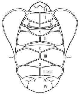
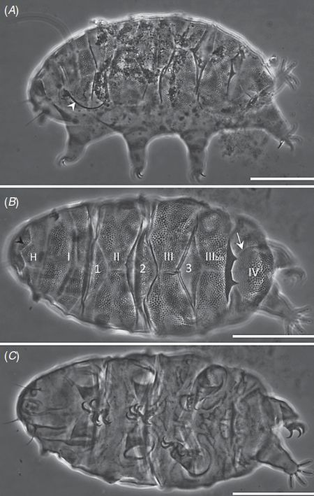
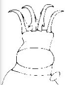
From Kristensen 1987: “Echiniscidae with black eyes: rigid buccal canal, stylet supports may be present, but very tiny and located close to the margin of pharyngeal bulb. All median plates present, plates 1 and 2 divided. Pseudosegmental plates IV’ paired or unpaired. Male with enlarged secondary clavae. Colour yellow or red. Emended description: The dorsal plates can be very difficult to see in some species, especially in the limnic forms. This must be considered as a secondary reduction. The head plate is large and may be deeply faceted or divided into smaller pieces. The small neck plate is not always visible. The segmental plate on the trunk consists of an unpaired scapular plate (I), a paired segmental plate II and III, and the caudal plate IV with terminal indentations. The pseudosegmental plate IV’ may be paired or unpaired. This character is highly variable, e.g. in the type species, P. suillus, some specimens have paired plates IV’, while other specimens from the same population have a single, unpaired plate. The same is true in the divided median plates 1 and 2; in nearly all species of Pseudechiniscus the plates are divided; this character may be overlooked if the preparation is not in very good condition. Median plate 3 is always undivided. Lateral segmental plates are present only in the more advanced species of Pseudechiniscus. The lateral plates are found only in connection with the trunk cirri B, C, D and E (P. victor-group) or lateral papillae (P. conifer-group). The lateral plates may be armed with a spine fringe. Leg plates are only well developed in species with trunk appendages. The plate of leg IV, especially, may have a dentated collar or spine fringe. Ventral plates are always absent. The cephalic cirri consist of papillate primary clavae, the cirri A, the papillate secondary clavae, external and internal cirri. The cirri can be split up in the tip as a tuft. The cirrus lack real cirrophore, but the base is slightly bulbous. The genus Pseudechiniscus consists of two groups, the suillus/conifer group without trunk cirri, and the victor group, where trunk cirri or spines are present. Leg sense organs are found on both leg I and IV. Typical tiny basal secondary spurs are located only on the internal claws. The buccal apparatus consists of a relatively long and rigid buccal tube, stylet supports are absent or reduced to thin fibres and very long stylets. The furcae of the stylets insert on the pharyngeal bulb. The armature of the pharyngeal bulb consists of bar-shaped placoids surrounded by thin cuticular lining.”

From Vecchi et al. 2016: “Echiniscid with pseudosegmental plate. Black eyes. Buccal cirri present, Filamentous cirri A present. Rigid buccal tube, stylet supports may be present, but very tiny and located close to the margin of pharyngeal bulb. Lateral and dorsal trunk filaments or spines generally absent or reduced in number and/or size. Notched collar (or spine fringe) on hind legs absent.”

Black eyes, no dentate collar.

Alberta:
none reported

Canada:
alberti Dastych, 1987
goedeni Grigarick, Mihelcic & Schuster, 1964
suillus (Ehrenberg, 1853) [Echiniscus mutabilis Murray, 1905]

North America / Nearctic:
brevimontanus Kendall-Fite & Nelson, 1996
n species 2

Other:
asper Abe, Utsugi & Takeda, 1998
bartkei Weglarska, 1962
beasleyi X. Li, L. Wang & Yu, 2007
bidenticulatus Bartoš, 1963
bispinosus (Murray, 1907)
clavatus Mihelcic, 1955
conifer (Richters, 1904)
dicrani Mihelcic, 1938
distinctus Mihelcic, 1951
facettalis Petersen, 1951 [Pseudechiniscus pseudoconifer facettalis Maucci, 1954] gullii Pilato & Lisi, 2006
insolitus Maucci, 1991
islandicus (Richters, 1904)
jiroveci Bartoš, 1963
juanitae de Barros, 1939 [Pseudechiniscus suillus franciscae de Barros, 1942] jubatus Biserov, 1990
megacephalus Mihelcic, 1951
nataliae Biserov & Maucci, 1986
novaezeelandiae novaezeelandiae (Richters, 1908)
novaezeelandiae aspinosa Iharos, 1963
novaezeelandiae laterospinosa Iharos, 1963
novaezeelandiae marinae Bartoš, 1934
occultus Dastych, 1980
papillosus X. Li, L. Wang, Liu & Su, 2005
pilatoi X. Li, 2007
pseudoconifer Ramazzotti, 1943
pulcher (Murray, 1910)
quadrilobatus Iharos, 1969
ramazzottii ramazzottii Maucci, 1952
ramazzottii facettalis Iharos, 1964 [Pseudechiniscus ramazzottii lineatus Maucci, 1973-74] raneyi Grigarick, Mihelcic & Schuster, 1964 (now multipseudechiniscus, Schulte & Miller, 2011
scorteccii Franceschi, 1952
shilinensis Yang, 2002
sinensis Rahm, 1937
spinerectus Pilato, Binda, Napolitano & Moncada, 2001
transsylvanicus Iharos, 1936
victor (Ehrenberg, 1853) [Pseudechiniscus tridentifer Bartoš, 1935] yunnanensis L. Wang, 2009


Citations :

Images from: Vecchi M, Cesari M, Bertolani R, Jönsson KI, Rebecchi L, Guidetti R. 2016. Integrative systematic studies on tardigrades from Antarctica identify new genera and new species within Macrobiotoidea. Invertebrate Systematics. 30: 303-322.

Claw image from Kristensen RM. 1987. Generic revision of the Echiniscidae (Heterotardigrada), with a discussion of the origin of the family. pp. 261-335 in Bertolani R (ed). Biology of Tardigrades: Selected symposia and monographs.

Ramazzotti G, Maucci W. 1983. Il phylum Tardigrada(III edizione riveduta e aggiornata). English translation by C. W. Beasley, 1995. Memorie dell’ Istituto Italiano di Idrobiologia 41: 1-1012.

Claw Symmetry Relative To Median Plane Of Leg?

Image from Bingemer J, Hohberg K. 2017. An illustrated identification key to the eutardigrade species (Tardigrada, Eutardigrada) presently known from European soils. Soil Organisms. 89 (3): 127-149.

Stylet Support Insertion Point As Percentage Of Buccal Tube Length

Stylet support insertion point = ss divided by Buccal tube length, as %. Note anterior measurements begin at anterior margin of stylet sheaths, ss is centred where stylet supports reach buccal tube (this requires a good dorsal or lateral view for proper measurement)


Image from Tumanov DV. 2006. Five new species of the genus Milnesium (Tardigrada, Eutardigrada, Milnesiidae). Zootaxa. 1122: 1-23.

Buccal Tube Long, Mouth At Anterior Of A Protrusible Snout

Images from Pilato G, Binda MG. 2010. Definition of families, subfamilies, genera, and subgenera of the Eutardigrada, and keys to their identification. Zootaxa. 2404: 1-54.

Pharyngeal Tube With Spiral Reinforcement

Images from Pilato G, Binda MG. 2010. Definition of families, subfamilies, genera, and subgenera of the Eutardigrada, and keys to their identification. Zootaxa. 2404: 1-54.

Peribuccal papillae?

Note: in a tardigrade with snout extended, the flex of the pharyngeal tube may not be obvious.


Images from Pilato G, Binda MG. 2010. Definition of families, subfamilies, genera, and subgenera of the Eutardigrada, and keys to their identification. Zootaxa. 2404: 1-54.

(Lateral) cephalic papillae, peribuccal papillae, Milnesiidae - type claws, exceptionally wide (and usually short) buccal tube?

Cephalic image modified from Nelson DR, Guidetti R, Rebecchi L. 2009. Tardigrada. Ch. 14 in Ecology and Classification of North American Freshwater Invertebrates, Thorp JH, Covich AP (eds.), 3rd ed.

Claw image modified from Pilato G, Binda MG. 2010. Definition of families, subfamilies, genera, and subgenera of the Eutardigrada, and keys to their identification. Zootaxa. 2404: 1-54.

Pharyngeal tube flexible?

Note: in a tardigrade with snout extended, the flex of the pharyngeal tube may not be obvious.


Images from Pilato G, Binda MG. 2010. Definition of families, subfamilies, genera, and subgenera of the Eutardigrada, and keys to their identification. Zootaxa. 2404: 1-54.

Serrated / dentate cuff / collar on legs IV

Left image from Kristensen RM. 1987. Generic revision of the Echiniscidae (Heterotardigrada), with a discussion of the origin of the family. pp. 261-335 in Bertolani R (ed). Biology of Tardigrades: Selected symposia and monographs.

Right Image from Richters F. 1926. Tardigrada. in Krumbach T. 1927. Handbuch der Zoologie, 3rd band, Walter de Gruyter & Co.

Leg sensory structures

Typically papilla on leg IV (se4 in image) and variable shape on leg I


Image from Kristensen RM. 1987. Generic revision of the Echiniscidae (Heterotardigrada), with a discussion of the origin of the family. pp. 261-335 in Bertolani R (ed). Biology of Tardigrades: Selected symposia and monographs.

Trunk appendages

Appendages (after Cirrus A) labeled according to plate … B (Scapular plate), C (First segmental plate), D (Second segmental plate), E (terminal plate). “B” is lateral, “Bd” is dorsal

Be careful! There can be great variability within a population, with individuals (especially juveniles) lacking some appendages. When in doubt, choose “Unclear” to ignore this character!


Left image from Ramazzotti G, Maucci W. 1983. Il phylum Tardigrada(III edizione riveduta e aggiornata). English translation by C. W. Beasley, 1995. Memorie dell’ Istituto Italiano di Idrobiologia 41: 1-1012.

Right image cropped from Kristensen RM. 1987. Generic revision of the Echiniscidae (Heterotardigrada), with a discussion of the origin of the family. pp. 261-335 in Bertolani R (ed). Biology of Tardigrades: Selected symposia and monographs.

Cirrus A form

Images modified from Kristensen RM. 1987. Generic revision of the Echiniscidae (Heterotardigrada), with a discussion of the origin of the family. pp. 261-335 in Bertolani R (ed). Biology of Tardigrades: Selected symposia and monographs.

Lower image (Mopsechiniscus) modified from du Bois-Reymond Marcus E. 1944. Sobre tardigrados brasileiros. Communicaciones Zoologicas del Museo de Historia Natural de Montevideo. 1(13): 1-19 plus plates.

Terminal / caudal plate with two notches?

Images modified from Kristensen RM. 1987. Generic revision of the Echiniscidae (Heterotardigrada), with a discussion of the origin of the family. pp. 261-335 in Bertolani R (ed). Biology of Tardigrades: Selected symposia and monographs.

First image (Echiniscus) modified from Richters F. 1926. Tardigrada. in Krumbach T. 1927. Handbuch der Zoologie, 3rd band, Walter de Gruyter & Co.

Inner & outer buccal cirri present

Image modified from Nelson DR, Guidetti R, Rebecchi L. 2009. Tardigrada. Ch. 14 in Ecology and Classification of North American Freshwater Invertebrates, Thorp JH, Covich AP (eds.), 3rd ed.

Pseudosegmental plate between last medial plate & terminal plate?

Note on 2nd image, what you see before the terminal plate is either no plate, or a full-width medial plate; don’t confuse the latter with a pseudosegmental plate!
Also, sometimes the pseudosegmental plate is paired, like segmental plates II & III. It’s still pseudosegmental!

Images from Clifford HF. 1991. Aquatic invertebrates of Alberta. University of Alberta Press, Alberta, Canada.

Segmental plates larger than intersegmental?

Image modified from Lindahl & Balser (1999), http://www.iwu.edu/~tardisdp/Keypage35.html

Any Paired Plates?

Images from Kristensen RM. 1987. Generic revision of the Echiniscidae (Heterotardigrada), with a discussion of the origin of the family. pp. 261-335