Echiniscidae

terrestrial species (found also in freshwater){Seminal receptacles absent. Dorsal plates present. Adult with 4 claws on each leg.}(Kristensen 1987) (Degma P, Guidetti R. 2018. Tardigrade Taxa. In: Water Bears: The biology of tardigrades. Schill RO, Editor. Switzerland: Springer Nature. p. 371-409. DOI: 10.1007/978-3-319-95702-9_15.)
Parechiniscus
From Ramazzotti & Maucci: “The plates are not well defined in the rostral part of the dorsum, better defined in the caudal part.” From Kristensen 1987: “Echiniscidae with small black eyes, rigid buccal tube, stylet supports absent, 8-9 dorsal plates, unpaired and more or less indistinct; secondary clavae dome shaped; uncoloured to grey. Emended description: […]
Mopsechiniscus
From Ramazzotti & Maucci 1983: “The plates are as in Pseudechiniscus; the internal and external buccal cirri are absent.” From Kristensen 1987: “Red Echiniscidae with black eyes, flexible buccal tube, stylet supports without CaCO3; ventral plates absent. Strongly sculptured dorsal plates. Paired pseudosegmental plates IV; all median plates undivided. A small rectangular plate with a […]
Cornechiniscus
From Ramazzotti & Maucci 1983 (tr. Beasley 1995): The plates are as in Pseudechiniscus; cirri A of conical shape, wide at the base, curved, internally hollow, with “saber blade” appearance. From Kristensen 1987: “Echiniscidae with black eyes, flexible buccal tube and paired pseudosegmental plates. Cirri A horn-shaped with a common base for the primary clavae, […]
Multipseudechiniscus
From Miller, Schulte, Johansson 2012: “Echiniscidae with dorsal segmental plates, pseudosegmental plates, and without ventral plates. Each segmental plate divided into two or more pieces, one or more piece paired. Anterior piece of cephalic and scapular plates paired. Posterior piece of trunk segment plates I, II, and III paired, forming pseudosegmental plates. Terminal plate divided […]
Acanthechiniscus
Acanthechiniscus, from Vecchi et al. 2016: “Echiniscid with pseudosegmental cuticular plate. Black eyes. Buccal cirri present, Filamentous cirri A present. Rigid buccal tube. Filaments and/or spines (may be small) present, generally in all lateral positions (B, C, D, E). Dorsal spines present. Notched collar (or spine fringe) on hind legs present.” Very similar to Pseudechiniscus, but notched cuff […]
Bryodelphax
From Ramazzotti & Maucci 1983 (tr. Beasley 1995): The median plates 1 and 2 are divided, not 3, so that there are visible total 5 median plates; lacking the notches on the terminal plate. From Kristensen 1987: “Sparsely pigmented Echiniscidae with red granulae eyes, unflexible buccal tube with CaCO3 encrusted stylet supports. Median plates 1 […]
Bryochoerus
From Ramazzotti & Maucci 1983 (tr. Beasley 1995): Median plates l, 2,3 divided transversely, so that total of 6 median plates visible. From Kristensen 1987: “Unpigmented to rose coloured Echiniscidae with red eyes, short inflexible buccal tube, with CaCO3 encrusted stylet supports. All median plates 1,2,3 divided, without pseudosegmental plates, with small lateral intersegmental plate […]
Testechiniscus
From Kristensen 1987: “Echiniscidae with black/brownish or ‘lipoid’ granular eyes; rigid buccal tube, large cuticular stylet supports, seven rows of sculptured ventral plates. Pseudosegmental plates absent. Paired segmental plates II and III with thinner anterior part. Caudal plate IV with a terminal indentation. Median plates 1 and 2 large, plate 3 small and reduced. Colour […]
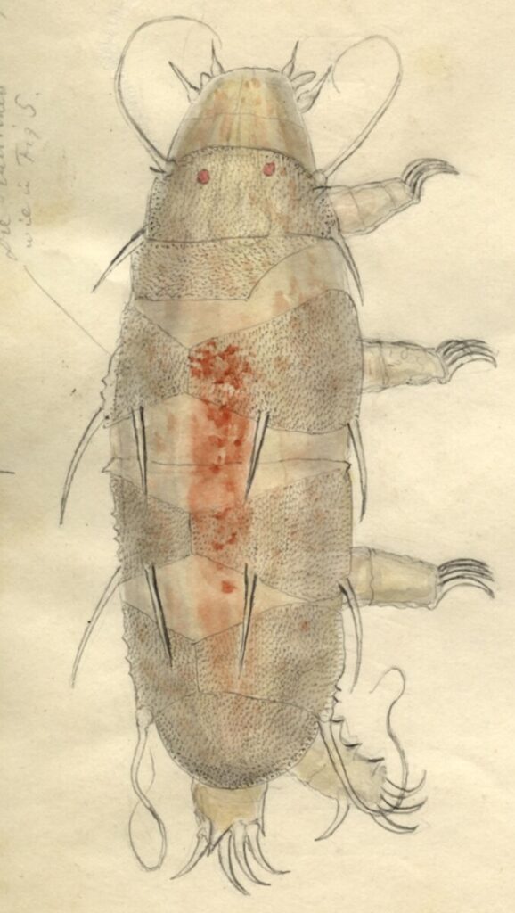
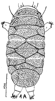
Hypechiniscus
From Thulin 1928: “Echiniscidae with armor containing five median plates. End plate adjacent to the last median plate. Terminal notches present. Echiniscus gladiator MURRAY var. exarmatus MURR leads to this genus. The exarmatus form (from the Faroe Islands) that I have examined differs in some aspects from MURRAYS description: The animals are colorless with black […]


