Mopsechiniscus

Image cropped from du Bois-Reymond Marcus E. 1944. Sobre tardigrados brasileiros. Communicaciones Zoologicas del Museo de Historia Natural de Montevideo. 1(13): 1-19 plus plates.

Class : Heterotardigrada

Order : Echiniscoidae

Family : Echiniscidae

Genus : Mopsechiniscus

From Ramazzotti & Maucci 1983: “The plates are as in Pseudechiniscus; the internal and external buccal cirri are absent.”

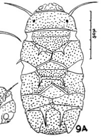
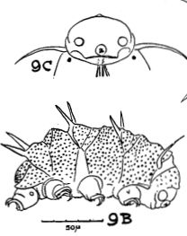
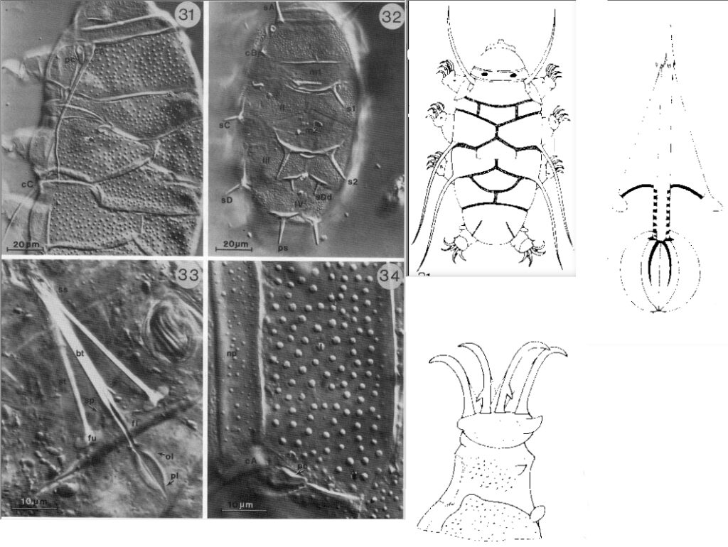
From Kristensen 1987: “Red Echiniscidae with black eyes, flexible buccal tube, stylet supports without CaCO3; ventral plates absent. Strongly sculptured dorsal plates. Paired pseudosegmental plates IV; all median plates undivided. A small rectangular plate with a «thorn» present on each side of the scapular plate (I). Cirrus A long stiff spine without cirrophore, primary clavae bent backwards. Internal and external cirri absent, secondary clavae small ovoid dome-shaped in female, large round dome-shaped in male. Emended description: The head bent strongly downwards; the mouth opening and the secondary clavae are located ventrally. The head plate is faceted with a small depression in the middle. The anterior margin of the head plate consists of two lateral ovoid structures. The neck plate is large and has the same sculpture as the head plate. This sculpture consists of coarse granulation and fine punctuation. The punctuation is formed by the pillar structure in the epicuticle. The coarse granulation consists of large, circular raised areas. The plates on the trunk segments have the same double granulation, but the circular raised areas are larger. The scapular plate (I) is relatively small with a rectangular plate on each. Cirri A and primary clavae are located between the scapular plate and the small lateral plates. Smaller lateral plates are also present in connection with the paired segmental plates II and III and the unpaired caudal plate. The large caudal plate has terminal indentation (notches). The posterior margin of the caudal plate is positioned latero-ventrally. The fourth pair of legs insert anterior to the margin of the plate. The median plates 1, 2 and 3 are undivided in adults, the median plate 2 is divided in the 2-clawed larva. Lateral intersegmental plates are present in connection with median plate 1. The reduction of the cephalic sense organs in the genus Mopsechiniscus is unique to the family Echiniscidae. The primary clavae previously overlooked (Marcus, 1929, Du Bois-Raymond Marcus, 1944) are best seen by Nomarski- optics. They are located in small depressions in the cuticle near the large base of the cirrus A. Each clava is bent backwards and adheres to the dorsal cuticle by a thin cuticular membrane. The cirrus A is a short spike in the larva, and a long thick spine in the adult. The cirri lack cirrophores, but each has a swollen base, which is continuous with the flagellum. The same type of cirrus is found on the trunk. The internal and external cirri are absent, the secondary clavae are dome-shaped and located ventrally. The collar or leg plates are absent on all legs, but a coarse granulation occurs on at least the fourth pairs of legs. Typical leg sense organs are located only on the fourth pair of legs. Furthermore, a triangular papilla is found on all legs, located near the base of the tarsus. The function of the latter is unknown, but it could be a muscle attachment. The tarsus is asymmetrical, with a cuticular cusp located near the base of the outer external claw, and a small cuticular cushion on the inner side of the tarsus. Each internal claw has a secondary spur. The 2-clawed larva of Mopsechiniscus has 8 pair of spines on the trunk. The four lateral spines are homologues with cirri B, C, D and E, but the two first pair of dorsal spines are located on the median plates 1 and 2, the third pair are small spine Dd and the fourth pair are pseudosegmental spines. At least two juvenile instars exist. The four-clawed juvenile lacking an anus, has reduced spine B, Dd and E, the four-clawed juvenile with an anus has only small ‘thorn’- shaped cirri B, C and a filiform cirri D. Of the dorsal appendages only the spines on median plate 2 and the pseudosegmental spine are present. The adult has filiform cirri C and D, a small thorn on the lateral plate could be a reduced cirri B. The pseudosegmental plates also have a pair of reduced triangular spines in a few animals. The buccal apparatus has a very long buccal tube. The anterior part of the tube is rigid, but, after the insertion of the very thin cuticular stylet supports, the tube becomes flexible, without CaCO3. The stylets are shorter than the buccal tube and do not reach the pharyngeal bulb. The furcae of the stylets are very large. The armature in the pharyngeal bulb consist of thick bar shaped placoids and a very thin outer cuticular lining.”

Image cropped from du Bois-Reymond Marcus E. 1944. Sobre tardigrados brasileiros. Communicaciones Zoologicas del Museo de Historia Natural de Montevideo. 1(13): 1-19 plus plates.
Images cropped from Kristensen RM. 1987. Generic revision of the Echiniscidae (Heterotardigrada), with a discussion of the origin of the family. pp. 261-335 in Bertolani R (ed). Biology of Tardigrades: Selected symposia and monographs.

Citations:

Kristensen RM. 1987. Generic revision of the Echiniscidae (Heterotardigrada), with a discussion of the origin of the family. pp. 261-335 in Bertolani R (ed). Biology of Tardigrades: Selected symposia and monographs.

Ramazzotti G, Maucci W. 1983. Il phylum Tardigrada(III edizione riveduta e aggiornata). English translation by C. W. Beasley, 1995. Memorie dell’ Istituto Italiano di Idrobiologia 41: 1-1012.

Claw Symmetry Relative To Median Plane Of Leg?

Image from Bingemer J, Hohberg K. 2017. An illustrated identification key to the eutardigrade species (Tardigrada, Eutardigrada) presently known from European soils. Soil Organisms. 89 (3): 127-149.

Stylet Support Insertion Point As Percentage Of Buccal Tube Length

Stylet support insertion point = ss divided by Buccal tube length, as %. Note anterior measurements begin at anterior margin of stylet sheaths, ss is centred where stylet supports reach buccal tube (this requires a good dorsal or lateral view for proper measurement)


Image from Tumanov DV. 2006. Five new species of the genus Milnesium (Tardigrada, Eutardigrada, Milnesiidae). Zootaxa. 1122: 1-23.

Buccal Tube Long, Mouth At Anterior Of A Protrusible Snout

Images from Pilato G, Binda MG. 2010. Definition of families, subfamilies, genera, and subgenera of the Eutardigrada, and keys to their identification. Zootaxa. 2404: 1-54.

Pharyngeal Tube With Spiral Reinforcement

Images from Pilato G, Binda MG. 2010. Definition of families, subfamilies, genera, and subgenera of the Eutardigrada, and keys to their identification. Zootaxa. 2404: 1-54.

Peribuccal papillae?

Note: in a tardigrade with snout extended, the flex of the pharyngeal tube may not be obvious.


Images from Pilato G, Binda MG. 2010. Definition of families, subfamilies, genera, and subgenera of the Eutardigrada, and keys to their identification. Zootaxa. 2404: 1-54.

(Lateral) cephalic papillae, peribuccal papillae, Milnesiidae - type claws, exceptionally wide (and usually short) buccal tube?

Cephalic image modified from Nelson DR, Guidetti R, Rebecchi L. 2009. Tardigrada. Ch. 14 in Ecology and Classification of North American Freshwater Invertebrates, Thorp JH, Covich AP (eds.), 3rd ed.

Claw image modified from Pilato G, Binda MG. 2010. Definition of families, subfamilies, genera, and subgenera of the Eutardigrada, and keys to their identification. Zootaxa. 2404: 1-54.

Pharyngeal tube flexible?

Note: in a tardigrade with snout extended, the flex of the pharyngeal tube may not be obvious.


Images from Pilato G, Binda MG. 2010. Definition of families, subfamilies, genera, and subgenera of the Eutardigrada, and keys to their identification. Zootaxa. 2404: 1-54.

Serrated / dentate cuff / collar on legs IV

Left image from Kristensen RM. 1987. Generic revision of the Echiniscidae (Heterotardigrada), with a discussion of the origin of the family. pp. 261-335 in Bertolani R (ed). Biology of Tardigrades: Selected symposia and monographs.

Right Image from Richters F. 1926. Tardigrada. in Krumbach T. 1927. Handbuch der Zoologie, 3rd band, Walter de Gruyter & Co.

Leg sensory structures

Typically papilla on leg IV (se4 in image) and variable shape on leg I


Image from Kristensen RM. 1987. Generic revision of the Echiniscidae (Heterotardigrada), with a discussion of the origin of the family. pp. 261-335 in Bertolani R (ed). Biology of Tardigrades: Selected symposia and monographs.

Trunk appendages

Appendages (after Cirrus A) labeled according to plate … B (Scapular plate), C (First segmental plate), D (Second segmental plate), E (terminal plate). “B” is lateral, “Bd” is dorsal

Be careful! There can be great variability within a population, with individuals (especially juveniles) lacking some appendages. When in doubt, choose “Unclear” to ignore this character!


Left image from Ramazzotti G, Maucci W. 1983. Il phylum Tardigrada(III edizione riveduta e aggiornata). English translation by C. W. Beasley, 1995. Memorie dell’ Istituto Italiano di Idrobiologia 41: 1-1012.

Right image cropped from Kristensen RM. 1987. Generic revision of the Echiniscidae (Heterotardigrada), with a discussion of the origin of the family. pp. 261-335 in Bertolani R (ed). Biology of Tardigrades: Selected symposia and monographs.

Cirrus A form

Images modified from Kristensen RM. 1987. Generic revision of the Echiniscidae (Heterotardigrada), with a discussion of the origin of the family. pp. 261-335 in Bertolani R (ed). Biology of Tardigrades: Selected symposia and monographs.

Lower image (Mopsechiniscus) modified from du Bois-Reymond Marcus E. 1944. Sobre tardigrados brasileiros. Communicaciones Zoologicas del Museo de Historia Natural de Montevideo. 1(13): 1-19 plus plates.

Terminal / caudal plate with two notches?

Images modified from Kristensen RM. 1987. Generic revision of the Echiniscidae (Heterotardigrada), with a discussion of the origin of the family. pp. 261-335 in Bertolani R (ed). Biology of Tardigrades: Selected symposia and monographs.

First image (Echiniscus) modified from Richters F. 1926. Tardigrada. in Krumbach T. 1927. Handbuch der Zoologie, 3rd band, Walter de Gruyter & Co.

Inner & outer buccal cirri present

Image modified from Nelson DR, Guidetti R, Rebecchi L. 2009. Tardigrada. Ch. 14 in Ecology and Classification of North American Freshwater Invertebrates, Thorp JH, Covich AP (eds.), 3rd ed.

Pseudosegmental plate between last medial plate & terminal plate?

Note on 2nd image, what you see before the terminal plate is either no plate, or a full-width medial plate; don’t confuse the latter with a pseudosegmental plate!
Also, sometimes the pseudosegmental plate is paired, like segmental plates II & III. It’s still pseudosegmental!

Images from Clifford HF. 1991. Aquatic invertebrates of Alberta. University of Alberta Press, Alberta, Canada.

Segmental plates larger than intersegmental?

Image modified from Lindahl & Balser (1999), http://www.iwu.edu/~tardisdp/Keypage35.html

Any Paired Plates?

Images from Kristensen RM. 1987. Generic revision of the Echiniscidae (Heterotardigrada), with a discussion of the origin of the family. pp. 261-335