Cornechiniscus

Kristensen RM. 1987. Generic revision of the Echiniscidae (Heterotardigrada), with a discussion of the origin of the family. pp. 261-335 in Bertolani R (ed). Biology of Tardigrades: Selected symposia and monographs.

Class : Heterotardigrada

Order : Echiniscoidae

Family : Echiniscidae

Genus : Cornechiniscus

From Ramazzotti & Maucci 1983 (tr. Beasley 1995): The plates are as in Pseudechiniscus; cirri A of conical shape, wide at the base, curved, internally hollow, with “saber blade” appearance.

From Kristensen 1987: “Echiniscidae with black eyes, flexible buccal tube and paired pseudosegmental plates. Cirri A horn-shaped with a common base for the primary clavae, secondary clavae ovoid, domed; internal and external cirri onion-shaped. Colour, yellow-red.

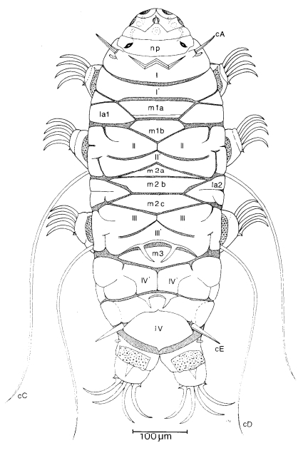
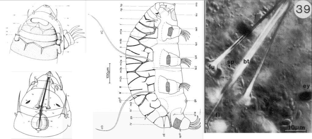
Emended description: One pair of subcephalic plates are located on the head and are finely sculptured. When the head is retracted, these plates function as a closing apparatus together with the anterior part of the dorsal head plate. The posterior part of the head plate has a stronger sculpture than the anterior part. Between the two sections there is a middorsal triangular structure in a zig- zag suture of very thin cuticle. The neck plate is very prominent and overlaps the posterior part of the head plate, when the head is retracted. The neck plate is very finely punctuated. The scapular plate (I) is divided into an anterior part with a very large W-shaped structure, and a posterior part. All after the different preparation methods the W-structure can be an elevation or a depression in the scapular plate, the posterior part can be totally separated from the anterior part of the scapular plate, as a pseudosegmental plate I’. In the same way, the segmental plates II and III each consist of an anterior paired piece and a nearly separated unpaired piece (pseudosegmental plates II’ and III’). The paired pseudosegmental plates IV’ may be nearly split up in small pieces by large indentations of both the anterior and posterior margins of the plates. The caudal plate (IV) may be divided into anterior and posterior parts or strongly notched. Unique for the genus Cornechiniscus is that the caudal plate does not cover the terminal part of the trunk segment IV; further- more, the fourth pair of legs are fused and located more terminally than in other Echiniscidae. The intersegmental plates are extremely well developed in Cor- nechiniscus, and a diagnostic character for this genus is a dorsal surface totally covered with sculptured plates except in the caudal region. The median plate 1 is divided in two pieces, the median plate 2 is divided in two or often in three, the median plate 3 is undivided. Lateral intersegmental plates are always pre- sent laterally to median plate 1 and 2, and can also be present in con- nection with median plate 3. Lateral bar-like structures are found on all four trunk segments; on segment II, III and IV the bar is found close to segmental plates, but on the first trunk segment (I) it is found in connection with the lateral intersegmental plate. Leg plates are present on all pairs of legs, on leg IV a long spine or smaller spines can be present in connection with the leg plate. The claws of leg IV are longer and curved less than those of the first three pairs of legs. One species (C. subcornutus) has a secondary spur on the external claws, other species have smooth claws. The head has short and horn shaped cirri A and primary clavae located be- tween the neck plate and the anterior part of the scapular plate. In most species these sense organs have their own plate and a common base for both the cirri and the clavae. The cirrus A has a distinct cirrophore, followed by a ball and socket link for the rigid flagellum. The flagellum has double cuticular wall with ridges. Cornechiniscus has the secondary clavae, external and internal cirri located latero-ventrally, as in the genus Mopsechiniscus. In both genera the head is bent strongly ventrally and the mouth opening is subterminal. The secon- dary clava is reduced to an ovoid dome, the internal cirrus (near the base of the internal cirrus, a so called «tertiary» clava can be present), is very short and onion-shaped with only a pointed tip. The external cirrus is longer and onion- shaped too, but has a short flagellum. Trunk cirri C and D are only present in one species, C. holmeni, dorsal spines can be found on segmental plates III and IV, as well as on the pseudosegmental plate IV’. Sense organs on the legs include a small spine on leg I and a small papilla on IV. The buccal tube is very long and bends between the head and the first trunk segment. The anterior part is rigid, but after the insertion of the stylet supports on the tube, it is flexible with a smaller diameter. The stylets are shorter than the buccal tube, the furca of the stylet never reaches the pharyngeal bulb. The cuticular stylet supports each have a small lens of CaCO3 encrusted material in- side. Posterior to the insertion of the stylet supports, the buccal canal is surrounded by cuticular amorphic material. In this same area large muscles attach, and thin cuticular attachments fibres support the thin flexible part of the buccal tube both anterior to and inside the pharyngeal bulb.”

Kristensen RM. 1987. Generic revision of the Echiniscidae (Heterotardigrada), with a discussion of the origin of the family. pp. 261-335 in Bertolani R (ed). Biology of Tardigrades: Selected symposia and monographs.

black eyes, flexible pt, paired pseudosegmental plates IV’

WARNING: IF CIRRUS A IS NOT DISTINCTLY DAGGER-LIKE (AND “HOLLOW”?), GO TO PSEUDECHINISCUS!


Citations:

Kristensen RM. 1987. Generic revision of the Echiniscidae (Heterotardigrada), with a discussion of the origin of the family. pp. 261-335 in Bertolani R (ed). Biology of Tardigrades: Selected symposia and monographs.

Ramazzotti G, Maucci W. 1983. Il phylum Tardigrada(III edizione riveduta e aggiornata). English translation by C. W. Beasley, 1995. Memorie dell’ Istituto Italiano di Idrobiologia 41: 1-1012.

Claw Symmetry Relative To Median Plane Of Leg?

Image from Bingemer J, Hohberg K. 2017. An illustrated identification key to the eutardigrade species (Tardigrada, Eutardigrada) presently known from European soils. Soil Organisms. 89 (3): 127-149.

Stylet Support Insertion Point As Percentage Of Buccal Tube Length

Stylet support insertion point = ss divided by Buccal tube length, as %. Note anterior measurements begin at anterior margin of stylet sheaths, ss is centred where stylet supports reach buccal tube (this requires a good dorsal or lateral view for proper measurement)


Image from Tumanov DV. 2006. Five new species of the genus Milnesium (Tardigrada, Eutardigrada, Milnesiidae). Zootaxa. 1122: 1-23.

Buccal Tube Long, Mouth At Anterior Of A Protrusible Snout

Images from Pilato G, Binda MG. 2010. Definition of families, subfamilies, genera, and subgenera of the Eutardigrada, and keys to their identification. Zootaxa. 2404: 1-54.

Pharyngeal Tube With Spiral Reinforcement

Images from Pilato G, Binda MG. 2010. Definition of families, subfamilies, genera, and subgenera of the Eutardigrada, and keys to their identification. Zootaxa. 2404: 1-54.

Peribuccal papillae?

Note: in a tardigrade with snout extended, the flex of the pharyngeal tube may not be obvious.


Images from Pilato G, Binda MG. 2010. Definition of families, subfamilies, genera, and subgenera of the Eutardigrada, and keys to their identification. Zootaxa. 2404: 1-54.

(Lateral) cephalic papillae, peribuccal papillae, Milnesiidae - type claws, exceptionally wide (and usually short) buccal tube?

Cephalic image modified from Nelson DR, Guidetti R, Rebecchi L. 2009. Tardigrada. Ch. 14 in Ecology and Classification of North American Freshwater Invertebrates, Thorp JH, Covich AP (eds.), 3rd ed.

Claw image modified from Pilato G, Binda MG. 2010. Definition of families, subfamilies, genera, and subgenera of the Eutardigrada, and keys to their identification. Zootaxa. 2404: 1-54.

Pharyngeal tube flexible?

Note: in a tardigrade with snout extended, the flex of the pharyngeal tube may not be obvious.


Images from Pilato G, Binda MG. 2010. Definition of families, subfamilies, genera, and subgenera of the Eutardigrada, and keys to their identification. Zootaxa. 2404: 1-54.

Serrated / dentate cuff / collar on legs IV

Left image from Kristensen RM. 1987. Generic revision of the Echiniscidae (Heterotardigrada), with a discussion of the origin of the family. pp. 261-335 in Bertolani R (ed). Biology of Tardigrades: Selected symposia and monographs.

Right Image from Richters F. 1926. Tardigrada. in Krumbach T. 1927. Handbuch der Zoologie, 3rd band, Walter de Gruyter & Co.

Leg sensory structures

Typically papilla on leg IV (se4 in image) and variable shape on leg I


Image from Kristensen RM. 1987. Generic revision of the Echiniscidae (Heterotardigrada), with a discussion of the origin of the family. pp. 261-335 in Bertolani R (ed). Biology of Tardigrades: Selected symposia and monographs.

Trunk appendages

Appendages (after Cirrus A) labeled according to plate … B (Scapular plate), C (First segmental plate), D (Second segmental plate), E (terminal plate). “B” is lateral, “Bd” is dorsal

Be careful! There can be great variability within a population, with individuals (especially juveniles) lacking some appendages. When in doubt, choose “Unclear” to ignore this character!


Left image from Ramazzotti G, Maucci W. 1983. Il phylum Tardigrada(III edizione riveduta e aggiornata). English translation by C. W. Beasley, 1995. Memorie dell’ Istituto Italiano di Idrobiologia 41: 1-1012.

Right image cropped from Kristensen RM. 1987. Generic revision of the Echiniscidae (Heterotardigrada), with a discussion of the origin of the family. pp. 261-335 in Bertolani R (ed). Biology of Tardigrades: Selected symposia and monographs.

Cirrus A form

Images modified from Kristensen RM. 1987. Generic revision of the Echiniscidae (Heterotardigrada), with a discussion of the origin of the family. pp. 261-335 in Bertolani R (ed). Biology of Tardigrades: Selected symposia and monographs.

Lower image (Mopsechiniscus) modified from du Bois-Reymond Marcus E. 1944. Sobre tardigrados brasileiros. Communicaciones Zoologicas del Museo de Historia Natural de Montevideo. 1(13): 1-19 plus plates.

Terminal / caudal plate with two notches?

Images modified from Kristensen RM. 1987. Generic revision of the Echiniscidae (Heterotardigrada), with a discussion of the origin of the family. pp. 261-335 in Bertolani R (ed). Biology of Tardigrades: Selected symposia and monographs.

First image (Echiniscus) modified from Richters F. 1926. Tardigrada. in Krumbach T. 1927. Handbuch der Zoologie, 3rd band, Walter de Gruyter & Co.

Inner & outer buccal cirri present

Image modified from Nelson DR, Guidetti R, Rebecchi L. 2009. Tardigrada. Ch. 14 in Ecology and Classification of North American Freshwater Invertebrates, Thorp JH, Covich AP (eds.), 3rd ed.

Pseudosegmental plate between last medial plate & terminal plate?

Note on 2nd image, what you see before the terminal plate is either no plate, or a full-width medial plate; don’t confuse the latter with a pseudosegmental plate!
Also, sometimes the pseudosegmental plate is paired, like segmental plates II & III. It’s still pseudosegmental!

Images from Clifford HF. 1991. Aquatic invertebrates of Alberta. University of Alberta Press, Alberta, Canada.

Segmental plates larger than intersegmental?

Image modified from Lindahl & Balser (1999), http://www.iwu.edu/~tardisdp/Keypage35.html

Any Paired Plates?

Images from Kristensen RM. 1987. Generic revision of the Echiniscidae (Heterotardigrada), with a discussion of the origin of the family. pp. 261-335