Aerobius

Aerobius habitus

Genus description abridged from Mapalo et al. 2004 (based on only extant specimen): “Body length is ~100 μm. The cuticle appears to be smooth with no observable protuberances. Cuticular folds are observed on the dorsal side, mostly likely due to its preservation in a shriveled state. Eyespots were not observed. A faint oval-shaped outline can […]
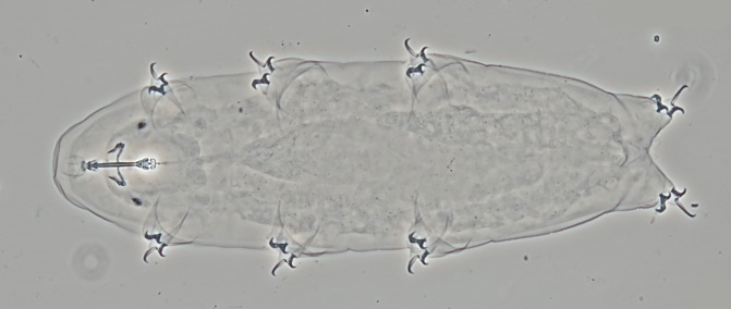
Parahypsibius

Genus description from Gąsiorek et al. 2024: “Hypsibiids with Ramazzottius-like claws and rigid buccal tube bent posteriorly. Only posterior bars may be present. AISMs asymmetrical, dorsal AISM shorter and higher than ventral, with prominent apex and less evident caudal processes. Pharynx with two granular macroplacoids. Rudimentary elliptical sensory organs present on head. Cuticular sculpturing well […]

Notahypsibius

Genus description from Tumanov 2020: “Hypsibiidae with Ramazzottius-like claws and completely rigid buccal tube. Apophyses for the insertion of the stylet muscles asymmetrical, dorsal AISM shorter and higher than ventral, with thickened anterior margin. Pharynx with two elongated macroplacoids and minute dot-like septulum. Cephalic elliptical organs present. Rugose cuticular sculpture. Eggs laid within the exuvium […]

Fontourion

Genus description from Gąsiorek et al. 2024: “Cuticle smooth. The AISMs are in the shape of semi-lunar hooks. Buccal tube is followed by a flexible annulated pharyngeal tube; the pharyngeal annulation is semi-complex (annuli single laterally and forking dorsoventrally). A DABT is present. There is a spherical pharynx, with two macroplacoids and a septulum. Macroplacoids […]

Degmion

Genus description from Gąsiorek et al. 2024: “Cuticle distinctly sculptured in the caudal portion of the trunk; more anterior portions with a weaker sculpturing or smooth. Apophyses for the insertion of the stylet muscles (AISMs) in the shape of semilunar hooks. Buccal tube followed by a flexible annulated pharyngeal tube; the pharyngeal annulation is semi-complex […]

Raribius

Genus description from Gąsiorek & Michalczyk 2020: “S-shaped stylet supports. Pharynx spherical, sometimes short and thin bar-like placoids present. Claws miniaturised, with external primary branches not markedly longer than secondary ones.” Species key: Gąsiorek P, Blagden B, Morek W, Michalczyk Ł. 2024. What is a ‘strong’synapomorphy? Redescriptions of Murray’s type species and descriptions of new […]

Insulobius

Genus description from Gąsiorek & Michalczyk 2020: “AISM reduced, in the shape of thin and flat ridges. Buccal tube followed by a flexible annulated pharyngeal tube, without the drop-like apodeme between them; the pharyngeal annulation type unknown. Pharyngeal tube annulation does not extend towards the more proximal portions of the buccal tube. Stylet supports and […]

Guidettion

Genus description from Gąsiorek & Michalczyk 2020: “Peribuccal structures absent. AISM in the shape of semilunar hooks. Buccal tube followed by a flexible annulated pharyngeal tube, without the drop-like apodeme between them; the pharyngeal annulation is simple (composed of single annuli). An ovoid pharynx with three bar-like, short or elongated or granular macroplacoids. Claws of […]

Arctodiphascon

Genus description from Tumanov & Tsvetkova 2023: “Peribuccal structures consisted of six peribuccal lobes. AISM in shape of “semilu­nar hooks” asymmetrical with respect to fron­tal plane with dorsal apophyses being distinctlyhigher, shorter and thicker than ventral ones, caudal processes of both apophyses pointing posteriorly and laterally. Buccal tube followed by a flexible annulated pharyngeal tube, […]

Cryobiotus

Genus description from Dastych 2019: “Median sized to large, dark-brown or blackish-brown pigmented hypsibiids with smooth cuticle (*) and large eyespots. Internal organs largely hidden by dense pigmentation. Six peribuccal lobes around mouth opening, peribuccal papillae not observed. The anterior apophyses of the mouth tube (= taa) of “acute hook” type (“Acutuncus variant”: Pilato & […]

Claw Symmetry Relative To Median Plane Of Leg?

Image from Bingemer J, Hohberg K. 2017. An illustrated identification key to the eutardigrade species (Tardigrada, Eutardigrada) presently known from European soils. Soil Organisms. 89 (3): 127-149.

Stylet Support Insertion Point As Percentage Of Buccal Tube Length

Stylet support insertion point = ss divided by Buccal tube length, as %. Note anterior measurements begin at anterior margin of stylet sheaths, ss is centred where stylet supports reach buccal tube (this requires a good dorsal or lateral view for proper measurement)


Image from Tumanov DV. 2006. Five new species of the genus Milnesium (Tardigrada, Eutardigrada, Milnesiidae). Zootaxa. 1122: 1-23.

Buccal Tube Long, Mouth At Anterior Of A Protrusible Snout

Images from Pilato G, Binda MG. 2010. Definition of families, subfamilies, genera, and subgenera of the Eutardigrada, and keys to their identification. Zootaxa. 2404: 1-54.

Pharyngeal Tube With Spiral Reinforcement

Images from Pilato G, Binda MG. 2010. Definition of families, subfamilies, genera, and subgenera of the Eutardigrada, and keys to their identification. Zootaxa. 2404: 1-54.

Peribuccal papillae?

Images from Pilato G, Binda MG. 2010. Definition of families, subfamilies, genera, and subgenera of the Eutardigrada, and keys to their identification. Zootaxa. 2404: 1-54.

(Lateral) cephalic papillae, peribuccal papillae, Milnesiidae - type claws, exceptionally wide (and usually short) buccal tube?

Cephalic image modified from Nelson DR, Guidetti R, Rebecchi L. 2009. Tardigrada. Ch. 14 in Ecology and Classification of North American Freshwater Invertebrates, Thorp JH, Covich AP (eds.), 3rd ed.

Claw image modified from Pilato G, Binda MG. 2010. Definition of families, subfamilies, genera, and subgenera of the Eutardigrada, and keys to their identification. Zootaxa. 2404: 1-54.

Pharyngeal tube flexible?

Note: in a tardigrade with snout extended, the flex of the pharyngeal tube may not be obvious.


Images from Pilato G, Binda MG. 2010. Definition of families, subfamilies, genera, and subgenera of the Eutardigrada, and keys to their identification. Zootaxa. 2404: 1-54.

Serrated / dentate cuff / collar on legs IV

Left image from Kristensen RM. 1987. Generic revision of the Echiniscidae (Heterotardigrada), with a discussion of the origin of the family. pp. 261-335 in Bertolani R (ed). Biology of Tardigrades: Selected symposia and monographs.

Right Image from Richters F. 1926. Tardigrada. in Krumbach T. 1927. Handbuch der Zoologie, 3rd band, Walter de Gruyter & Co.

Leg sensory structures

Typically papilla on leg IV (se4 in image) and variable shape on leg I


Image from Kristensen RM. 1987. Generic revision of the Echiniscidae (Heterotardigrada), with a discussion of the origin of the family. pp. 261-335 in Bertolani R (ed). Biology of Tardigrades: Selected symposia and monographs.

Trunk appendages

Appendages (after Cirrus A) labeled according to plate … B (Scapular plate), C (First segmental plate), D (Second segmental plate), E (terminal plate). “B” is lateral, “Bd” is dorsal

Be careful! There can be great variability within a population, with individuals (especially juveniles) lacking some appendages. When in doubt, focus on other characters first!


Left image from Ramazzotti G, Maucci W. 1983. Il phylum Tardigrada(III edizione riveduta e aggiornata). English translation by C. W. Beasley, 1995. Memorie dell’ Istituto Italiano di Idrobiologia 41: 1-1012.

Right image cropped from Kristensen RM. 1987. Generic revision of the Echiniscidae (Heterotardigrada), with a discussion of the origin of the family. pp. 261-335 in Bertolani R (ed). Biology of Tardigrades: Selected symposia and monographs.

Cirrus A form

Images modified from Kristensen RM. 1987. Generic revision of the Echiniscidae (Heterotardigrada), with a discussion of the origin of the family. pp. 261-335 in Bertolani R (ed). Biology of Tardigrades: Selected symposia and monographs.

Lower image (Mopsechiniscus) modified from du Bois-Reymond Marcus E. 1944. Sobre tardigrados brasileiros. Communicaciones Zoologicas del Museo de Historia Natural de Montevideo. 1(13): 1-19 plus plates.

Terminal / caudal plate with two notches?

Images modified from Kristensen RM. 1987. Generic revision of the Echiniscidae (Heterotardigrada), with a discussion of the origin of the family. pp. 261-335 in Bertolani R (ed). Biology of Tardigrades: Selected symposia and monographs.

First image (Echiniscus) modified from Richters F. 1926. Tardigrada. in Krumbach T. 1927. Handbuch der Zoologie, 3rd band, Walter de Gruyter & Co.

Inner & outer buccal cirri present

Image modified from Nelson DR, Guidetti R, Rebecchi L. 2009. Tardigrada. Ch. 14 in Ecology and Classification of North American Freshwater Invertebrates, Thorp JH, Covich AP (eds.), 3rd ed.

Pseudosegmental plate between last medial plate & terminal plate?

Note on 2nd image, what you see before the terminal plate is either no plate, or a full-width medial plate; don’t confuse the latter with a pseudosegmental plate!
Also, sometimes the pseudosegmental plate is paired, like segmental plates II & III. It’s still pseudosegmental!

Images from Clifford HF. 1991. Aquatic invertebrates of Alberta. University of Alberta Press, Alberta, Canada.

Segmental plates larger than intersegmental?

Image modified from Lindahl & Balser (1999), http://www.iwu.edu/~tardisdp/Keypage35.html

Any Paired Plates?

Modified from Lindahl K, Balser S.  1999. Key to tardigrade genera [Internet]. Available from: https://sun.iwu.edu/~tardisdp/Keypage33.html