Platicrista

Marley NJ. 2006. A new species of Tardigrada, Platicrista ramsayi sp. nov. from the páramo of volcán Chiles, Ecuador. Zootaxa. 1166: 35-48.

Class : Eutardigrada

Order : Hypsibioidea

Family : Hypsibiidae

N/A

Hypsibioidea from Pilato 1969 in Marley et al. 2011: “Parachela; claws asymmetrical (2121); Hypsibius-type claw pairs; AISM hooked (or, if the buccal tube is elongated, AISM can be broad ridges).”

Hypsibioidea from Bertolani et al. 2014: “Double claws asymmetrical with respect to the median plane of the leg (2121), the external (or posterior) claw often with flexible main branch; double claws different in size and shape on the same leg (Hypsibius and Ramazzottius type, or modified), or very reduced in size (Calohypsibius and Microhypsibius type); buccal tube often very narrow”

Hypsibioidea from Gąsiorek et al. 2018: “Eutardigrades with asymmetrical claws (2-1-2-1) and pseudolunulae at claw bases or without any cuticular structures under the basal parts. Hooked or broad-ridged apophyses for the insertion of the stylet muscles. Herbivorous or microbivorous (Guidetti et al. 2012).”

Hypsibiidae from Pilato 1969: “Senza alcuna appendice cefalica. Le due diplounghie di ciascuna zampa sono disposte asimmetricamente rispetto al piano mediano della zampa stessa e posssono essere di tipo Isohypsibius o di tipo Hypsibius. In ogni zampa le due diplounghie sono talvolta quasi uguali fra loro ma molto spesso profondamente diverse per forma e dimensioni. Lunule più spesso assenti, presenti in poche specie. L’apparato boccale è generalmente di tipo Hypsibius o Diphascon e soltanto nel genere Doryphoribius è di tipo Macrobiotus. Il bulbo faringeo è generalmente provvisto di placoidi, ne è soltanto nel genere Itaquascon.”

Translated: Without any cephalic appendages. The two double-claws of each leg are arranged asymmetrically with respect to the median plane of the leg itself and can be of the Isohypsibius or Hypsibius type. In each paw the two double-claws are sometimes almost equal to each other but very often very different in shape and size. Lunule more often absent, present in few species. The buccal apparatus is generally of the Hypsibius or Diphascon type and only in the genus Doryphoribius is of the Macrobiotus type. The pharyngeal bulb is generally provided with placoids, except in the genus Itaquascon.

Hypsibiidae from Bertolani et al. 2014: “Double claws different in shape and size on the same leg, the external (or posterior) of Hypsibius type claw with the secondary branch forming a continuous hook with its basal tract and the primary branch connected to the basal tract with a flexible part. Septulum present in the pharynx of some genera. Eggs smooth (but rarely weakly ornamented) laid within the exuvium, in some cases eggs ornamented laid free.”

Hypsibiidae from Gąsiorek et al. 2018: “Eutardigrades without cephalic papillae (sensu structures present e.g. in Halobiotus Kristensen, 1982; see Møbjerg et al. 2007) and elliptical organs. Claws of the Hypsibius type, i.e. asymmetrical both with respect to the sequence of primary and secondary branches (2-1-2-1) and with respect to the size, with external and posterior claws being always clearly larger than internal and anterior claws. Accessory points symmetrical. Two types of bucco-pharyngeal apparatuses: with the buccal tube rigid over its entire length (Hypsibiinae) or with a rigid anterior buccal tube followed by a flexible posterior pharyngeal tube (all remaining subfamilies).”

Itaquasconinae from Pilato 1969: “Unghie di tipo Hypsibius; apparato boccale di tipo Diphascon; placoidi presenti o assenti.”

Translated: Claws of Hypsibius type; buccal apparatus of Diphascon type; placoids present or absent.

Itaquasconinae from Bertolani et al. 2014: “Buccal tube followed by a flexible portion, without cuticular thickening between them; the flexible portion is an annulated pharyngeal tube in all genera (Parascon excluded); pharyngeal bulb very elongated; placoids very long and in line, sometime reduced to a bulbar lining.”

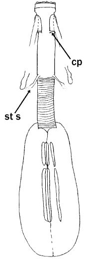
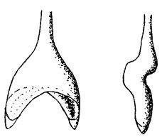
Genus description from Pilato 1987: “The bucco-pharyngeal apparatus is of the Diphascon model with the apophyses for the insertion of the muscles of the stylets in the shape of ‘very wide and flat ridges’; they are symmetrical with respect to the frontal plane; and with the caudal processes poorly developed and pointing sideways; the furcae have the postero-lateral processes spoon-like and tapering at their apices. Other characters: the bucco-pharyngeal tube is very wide and it lacks the ‘drop-like’ thickening; the pharyngeal tube is as long as the bulb and as long as the buccal tube or slightly shorter; the stylet supports are inserted at the very end of the buccal tube; the pharyngeal bulb is very lengthened and so are the placoids; the pharyngeal apophyses are absent.”

Pilato G, Binda MG. 2010. Definition of families, subfamilies, genera, and subgenera of the Eutardigrada, and keys to their identification. Zootaxa. 2404: 1-54.

Citations:

Bertolani R, Guidetti R, Marchioro T, Altiero T, Rebecchi L, Cesari M. 2014. Phyloeny of Eutardigrada: New molecular data and their morphological support lead to the identification of new evolutionary lineages. Molecular Phylogenetics and Evolution. 76: 110-126.

Gąsiorek P, Stec D, Morek W, Michalczyk Ł. 2018. An integrative redescription of Hypsibius dujardini (Doyère, 1840), the nominal taxon for Hypsibioidea (Tardigrada: Eutardigrada). Zootaxa. 4415 (1): 45-75.

Marley NJ, McInnes SJ, Sands CJ. 2011. Phylum Tardigrada: A re-evaluation of the Parachela. Zootaxa. 2819: 51-64.

Pilato G. 1969. Schema per una nuova sistemazione delle famiglie e dei generi degli Eutardigrada. Bollettino delle Sedute della Accademia Gioenia di Scienze Naturali in Catania, Series IV. 10 (277): 181-193.

Pilato G. 1987. Revision of the genus Diphascon Plate, 1889, with remarks on the subfamily Itaquasconinae (Eutardigrada, Hypsibiidae). pp. 337-357 in Bertolani R (ed). Biology of Tardigrades: Selected symposia and monographs.

Claw Symmetry Relative To Median Plane Of Leg?

Image from Bingemer J, Hohberg K. 2017. An illustrated identification key to the eutardigrade species (Tardigrada, Eutardigrada) presently known from European soils. Soil Organisms. 89 (3): 127-149.

Stylet Support Insertion Point As Percentage Of Buccal Tube Length

Stylet support insertion point = ss divided by Buccal tube length, as %. Note anterior measurements begin at anterior margin of stylet sheaths, ss is centred where stylet supports reach buccal tube (this requires a good dorsal or lateral view for proper measurement)


Image from Tumanov DV. 2006. Five new species of the genus Milnesium (Tardigrada, Eutardigrada, Milnesiidae). Zootaxa. 1122: 1-23.

Buccal Tube Long, Mouth At Anterior Of A Protrusible Snout

Images from Pilato G, Binda MG. 2010. Definition of families, subfamilies, genera, and subgenera of the Eutardigrada, and keys to their identification. Zootaxa. 2404: 1-54.

Pharyngeal Tube With Spiral Reinforcement

Images from Pilato G, Binda MG. 2010. Definition of families, subfamilies, genera, and subgenera of the Eutardigrada, and keys to their identification. Zootaxa. 2404: 1-54.

Peribuccal papillae?

Note: in a tardigrade with snout extended, the flex of the pharyngeal tube may not be obvious.


Images from Pilato G, Binda MG. 2010. Definition of families, subfamilies, genera, and subgenera of the Eutardigrada, and keys to their identification. Zootaxa. 2404: 1-54.

(Lateral) cephalic papillae, peribuccal papillae, Milnesiidae - type claws, exceptionally wide (and usually short) buccal tube?

Cephalic image modified from Nelson DR, Guidetti R, Rebecchi L. 2009. Tardigrada. Ch. 14 in Ecology and Classification of North American Freshwater Invertebrates, Thorp JH, Covich AP (eds.), 3rd ed.

Claw image modified from Pilato G, Binda MG. 2010. Definition of families, subfamilies, genera, and subgenera of the Eutardigrada, and keys to their identification. Zootaxa. 2404: 1-54.

Pharyngeal tube flexible?

Note: in a tardigrade with snout extended, the flex of the pharyngeal tube may not be obvious.


Images from Pilato G, Binda MG. 2010. Definition of families, subfamilies, genera, and subgenera of the Eutardigrada, and keys to their identification. Zootaxa. 2404: 1-54.

Serrated / dentate cuff / collar on legs IV

Left image from Kristensen RM. 1987. Generic revision of the Echiniscidae (Heterotardigrada), with a discussion of the origin of the family. pp. 261-335 in Bertolani R (ed). Biology of Tardigrades: Selected symposia and monographs.

Right Image from Richters F. 1926. Tardigrada. in Krumbach T. 1927. Handbuch der Zoologie, 3rd band, Walter de Gruyter & Co.

Leg sensory structures

Typically papilla on leg IV (se4 in image) and variable shape on leg I


Image from Kristensen RM. 1987. Generic revision of the Echiniscidae (Heterotardigrada), with a discussion of the origin of the family. pp. 261-335 in Bertolani R (ed). Biology of Tardigrades: Selected symposia and monographs.

Trunk appendages

Appendages (after Cirrus A) labeled according to plate … B (Scapular plate), C (First segmental plate), D (Second segmental plate), E (terminal plate). “B” is lateral, “Bd” is dorsal

Be careful! There can be great variability within a population, with individuals (especially juveniles) lacking some appendages. When in doubt, choose “Unclear” to ignore this character!


Left image from Ramazzotti G, Maucci W. 1983. Il phylum Tardigrada(III edizione riveduta e aggiornata). English translation by C. W. Beasley, 1995. Memorie dell’ Istituto Italiano di Idrobiologia 41: 1-1012.

Right image cropped from Kristensen RM. 1987. Generic revision of the Echiniscidae (Heterotardigrada), with a discussion of the origin of the family. pp. 261-335 in Bertolani R (ed). Biology of Tardigrades: Selected symposia and monographs.

Cirrus A form

Images modified from Kristensen RM. 1987. Generic revision of the Echiniscidae (Heterotardigrada), with a discussion of the origin of the family. pp. 261-335 in Bertolani R (ed). Biology of Tardigrades: Selected symposia and monographs.

Lower image (Mopsechiniscus) modified from du Bois-Reymond Marcus E. 1944. Sobre tardigrados brasileiros. Communicaciones Zoologicas del Museo de Historia Natural de Montevideo. 1(13): 1-19 plus plates.

Terminal / caudal plate with two notches?

Images modified from Kristensen RM. 1987. Generic revision of the Echiniscidae (Heterotardigrada), with a discussion of the origin of the family. pp. 261-335 in Bertolani R (ed). Biology of Tardigrades: Selected symposia and monographs.

First image (Echiniscus) modified from Richters F. 1926. Tardigrada. in Krumbach T. 1927. Handbuch der Zoologie, 3rd band, Walter de Gruyter & Co.

Inner & outer buccal cirri present

Image modified from Nelson DR, Guidetti R, Rebecchi L. 2009. Tardigrada. Ch. 14 in Ecology and Classification of North American Freshwater Invertebrates, Thorp JH, Covich AP (eds.), 3rd ed.

Pseudosegmental plate between last medial plate & terminal plate?

Note on 2nd image, what you see before the terminal plate is either no plate, or a full-width medial plate; don’t confuse the latter with a pseudosegmental plate!
Also, sometimes the pseudosegmental plate is paired, like segmental plates II & III. It’s still pseudosegmental!

Images from Clifford HF. 1991. Aquatic invertebrates of Alberta. University of Alberta Press, Alberta, Canada.

Segmental plates larger than intersegmental?

Image modified from Lindahl & Balser (1999), http://www.iwu.edu/~tardisdp/Keypage35.html

Any Paired Plates?

Images from Kristensen RM. 1987. Generic revision of the Echiniscidae (Heterotardigrada), with a discussion of the origin of the family. pp. 261-335