Richtersiusidae

Class: Eutardigrada

Order: Parachela

Superfamily: Macrobiotoidea

Click taxa for descriptions

Richtersiidae from Guidetti et al. 2016: “Double claws Y-shaped, with the two branches forming an evident common tract of variable length. Large teeth on all lunules. Buccal tube with ventral lamina and a cuticular thick on the anterior, dorsal wall of the buccal tube (which can form a large apophysis). Absence of transverse crests in the buccal armature. Two macroplacoids in the pharynx. Cuticular pores (at least in a phase of the life cycle). Eggs laid freely with cuticular processes on their surface.”

Richtersiidae from Lisi et al. 2020: “Presence of cuticular pores at least in some life stages. Buccal tube with ventral lamina provided with an additional anterior ventral thickening which can form a large apophysis in some genera; a cuticular thickening on the anterior, dorsal wall of the buccal tube sometimes developed into a dorsal apophysis. Two macroplacoids in the pharynx. Symmetric double claws Y-shaped, with stalk system and the two branches forming an evident common tract of variable length. Large teeth on all lunules. Eggs laid freely with cuticular processes on their surface.”

Richtersiusidae from Guidetti et al. 2021: “Double claws Y-shaped, with the two branches forming an evident common tract of variable length. Large teeth on all lunules. Buccal tube with ventral lamina and a cuticular thickness on the anterior portion of the dorsal wall of the buccal tube (which can form a large apophysis). Two macroplacoids in the pharynx. Cuticular pores (at least in one phase of the life cycle). Eggs laid freely with processes on their surface.”

Richtersiusidae from Stec 2023: “Double claws Y-shaped, with the two branches forming an evident common tract of a variable length with system of internal septa. In majority of taxa included within the family, teeth present in lunulae on all legs. Buccal tube with ventral lamina exhibiting ventral thickening in its anterior portion (sometimes hardly visible under light microcope) and a cuticular thick on the anterior, dorsal wall of the buccal tube (which can form a large apophysis). Absence of transverse crests in the buccal armature. Two macroplacoids in the pharynx. Microplacoid absent. Cuticular pores (at least in a phase of the life cycle). Eggs laid freely with conical (usually spiky) processes and without areolation on their surface. Body and leg granulation absent in all currently recognized species.”

Richtersiusidae from Massa et al. 2024: “Double claws Y-shaped, with the two branches forming an evident common tract of a variable length with system of internal septa. Teeth present or absent on lunulae I-III and always present on lunulae IV. Buccal tube with ventral lamina exhibiting ventral thickening in its anterior portion
(sometimes hardly visible under light microscope) and a cuticular thickening on the anterior, dorsal wall of the buccal tube (which can form a large apophysis). Transverse crests in the buccal armature absent. Two macroplacoids in the pharynx. Microplacoid present or absent (in most taxa). Cuticular pores (at least in a phase of the life cycle). Eggs laid freely with conical (usually spiky) processes and without areolation on their surface.
Body and leg granulation absent in all currently recognized species.”


Citations:

Guidetti, R., Rebecchi, L., Bertolani, R., Jönsson, K.I., Kristensen, R.M. & Cesari, M. (2016) Morphological and molecular analyses on Richtersius (Eutardigrada) diversity reveal its new systematic position and lead to the establishment of a new genus and a new family within Macrobiotoidea. Zoological Journal of the Linnean Society, 178, 834–845.

Guidetti, R., Schill, R.O., Giovannini, I., Massa, E., Goldoni, S.E., Ebel, Ch., Förschler, M.I., Rebecchi, L. & Cesari, M. (2021) When DNA sequence data and morphological results fit together: Phylogenetic position of Crenubiotus within Macrobiotoidea (Eutardigrada) with description of Crenubiotus ruhesteini sp. nov. Journal of Zoological Systematics and Evolutionary Research. https://doi.org/10.1111/jzs.12449, 59 (3), 576–587.

Lisi, O., Londoño, R. & Quiroga, S. (2020) Description of a new genus and species (Eutardigrada: Richtersiidae) from Colombia, with comments on the family Richtersiidae. Zootaxa, 4822 (4): 531–550.

Massa, E., Vecchi, M., Calhim, S. & Choong, H. (2024) First records of limnoterrestrial tardigrades (Tardigrada) from Haida Gwaii, British Columbia, Canada. The European Zoological Journal, 91, 1–20. https://doi.org/10.1080/24750263.2023.2288824

Stec, D. (2023) Integrative taxonomy helps to revise systematics and questions the purported cosmopolitan nature of the type species within the genus Diaforobiotus (Eutardigrada: Richtersiusidae). Organisms Diversity & Evolution, 23 (2), 309–328. https://doi.org/10.1007/s13127-022-00592-6
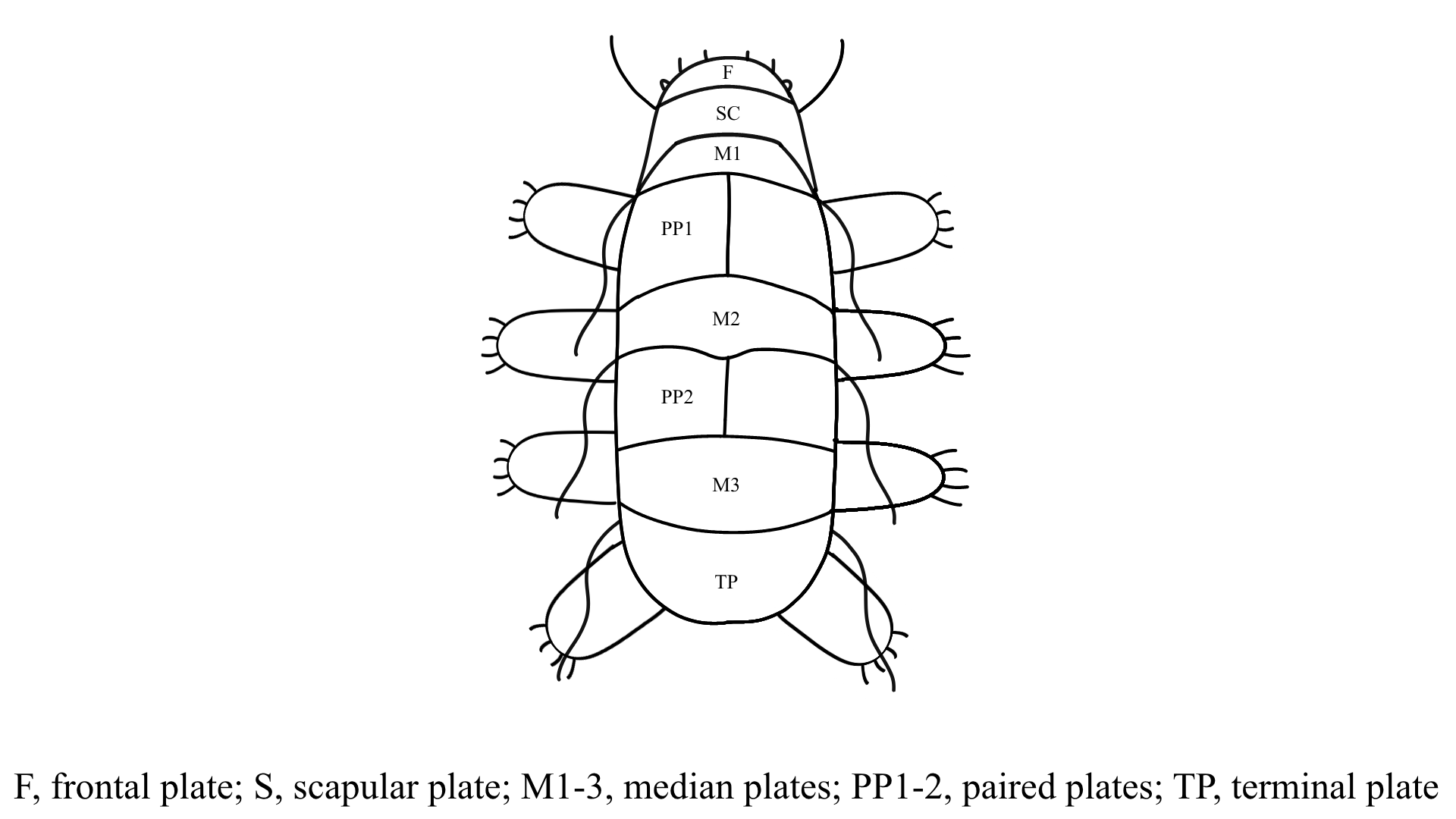
Armoured (dorsal) plates?

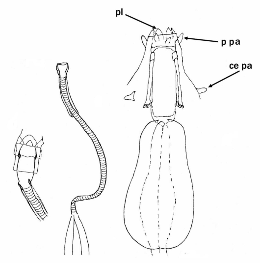
Pharyngeal tube flexible?

Note: in a tardigrade with snout extended, the flex of the pharyngeal tube may not be obvious.


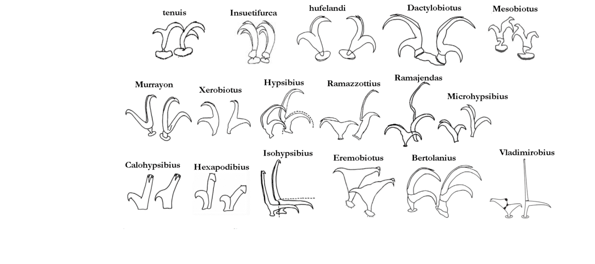
Images from Pilato G, Binda MG. 2010. Definition of families, subfamilies, genera, and subgenera of the Eutardigrada, and keys to their identification. Zootaxa. 2404: 1-54.

Claw type

tenuis: With stalk, a long common tract formed by the joined primary & secondary branches, and distal sections of both the secondary branch – shorter than the primary – and primary branch forming almost a right angle.
Insuetifurca: Modified hufelandi: Basal section subdivided into a very short stem & a large distal portion.
hufelandi: Primary & secondary branches rigidly joined, forming an acute angle, with variable but long common basal tract and cylindrical stalk.
Dactylobiotus: Basal section a trapezoidal lamina; secondary branch clearly shorter than primary and inserted near the base of the latter; the two branches form an almost right angle.
Mesobiotus: Primary & secondary branches rigidly joined, forming acute angle, with variable but long common basal tract with internal septum defining a distal part, with cylindrical stalk. Often very prominent, strong accessory point on primary.
Murrayon: Basal section a trapezoidal lamina; primary & secondary branches joined to each other for a very short portion and form an acute angle.
Xerobiotus: Whole double claw equally sclerified; basal section not subdivided into a basal stem & distinct distal portion; no separating septa present; primary & secondary branches rigidly joined forming an acute angle.
Hypsibius: Secondary branch forming a continuous curve with its basal tract; in external claws the primary branch connected to the basal tract with a flexible part while the inner claw is rigid.
Ramazzottius: External claws with basal section longer than the secondary branch; primary branch very long and slender and connected to basal tract with an evident, thin, flexible tract; internal claws short & stout, rigid.
Ramajendas: External claws similar to Hypsibius type with extremely long and slender primary branch; internal claw similar to Isohypsibius type.
Microhypsibius: Small, rigid, with an evident thin basal tract continuous with the primary branch; secondary branch rigidly joined to the primary branch at a distance from the base of the claw.
Calohypsibius: Small, rigid, in frontal view with a base as large as the sum of the primary and secondary branch widths, without a suture between the two branches.
Hexapodibius: Very short, without common basal tract, with base as large as the sum of the primary and secondary branches.
Isohypsibius: Secondary branch inserted perpendicularly on the claw basal tract.
Eremobiotus: Isohypsibius-type very modified with two branches of each claw joined to one another for a long portion of their length constituting a large common tract from which the two distal tracts diverge forming an angle of almost 180°.
Bertolanius: Subdivided into three distinct sections: basal section, secondary branch and primary branch (rigidly joined to the secondary branch), one on top of the other and separated by septa; the angles between the main and secondary branch different in external and internal claws – and acute angle (about 45°) is formed by the external claw, and an almost right angle (about 80°) by the internal claw; internal claws can rotate on their base up to 180°.
Vladimirobius: external at least 2x length of internal (measured from base of claw), internal very wide at the junction of primary and secondary branches, short and thin basal part, primary branches with very prominent gibbosity-like projection near the junction of primary and secondary branches; external very thin with long basal part; accessory points of both claws very near to end of primary branch.
Calcarobiotus: Basal section, with or without basal spurs, subdivided into a thin flexible stem & a wide distal section in the shape of an upside-down triangle distally delmited by a septum; primary & secondary branches similar in shape & size.
Macroversum: Basal section subdivided into a thin, flexible, stem and distal portion not very sclerotized; primary & secondary branches connected to each other for a short portion and form an almost right angle.

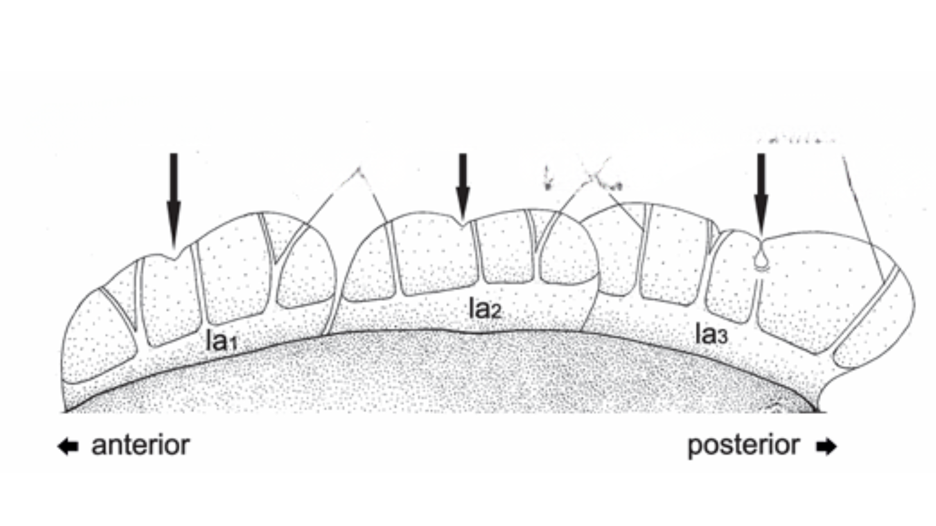
Number of lateral alae

Hansen, J.G. & Kristensen, R.M. (2021) A new genus and five new species of the subfamily Florarctinae (Tardigrada, Arthrotardigrada). European Journal of Taxonomy, 762, 149-184.

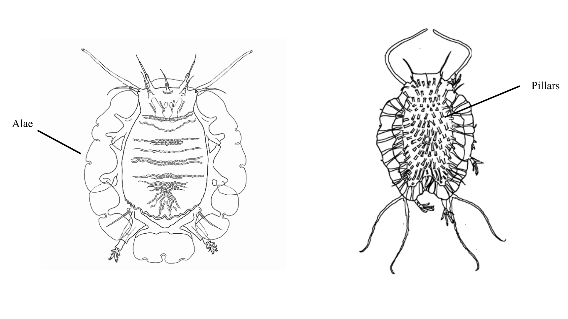
Epicuticle

Left image: Anguas-Escalante, A., De Jesús Navarrete, A., Demilio, E., Pérez-Pech, W.A. & Hansen, J.G. (2020). A new species of Tardigrada from a Caribbean reef lagoon, Florarctus yucatanensis sp. nov. (Halechiniscidae: Florarctinae). Cahiers de Biologie Marine, 62, 377-385.

Right image: Fontoura, P., Bartels, B.J., Jørgensen, A., Kristensen, R.M. & Hansen, J.G. (2017) A dichotomous key to the genera of the marine Heterotardigrades (Tardigrada). Zootaxa, 4294, 1-45.

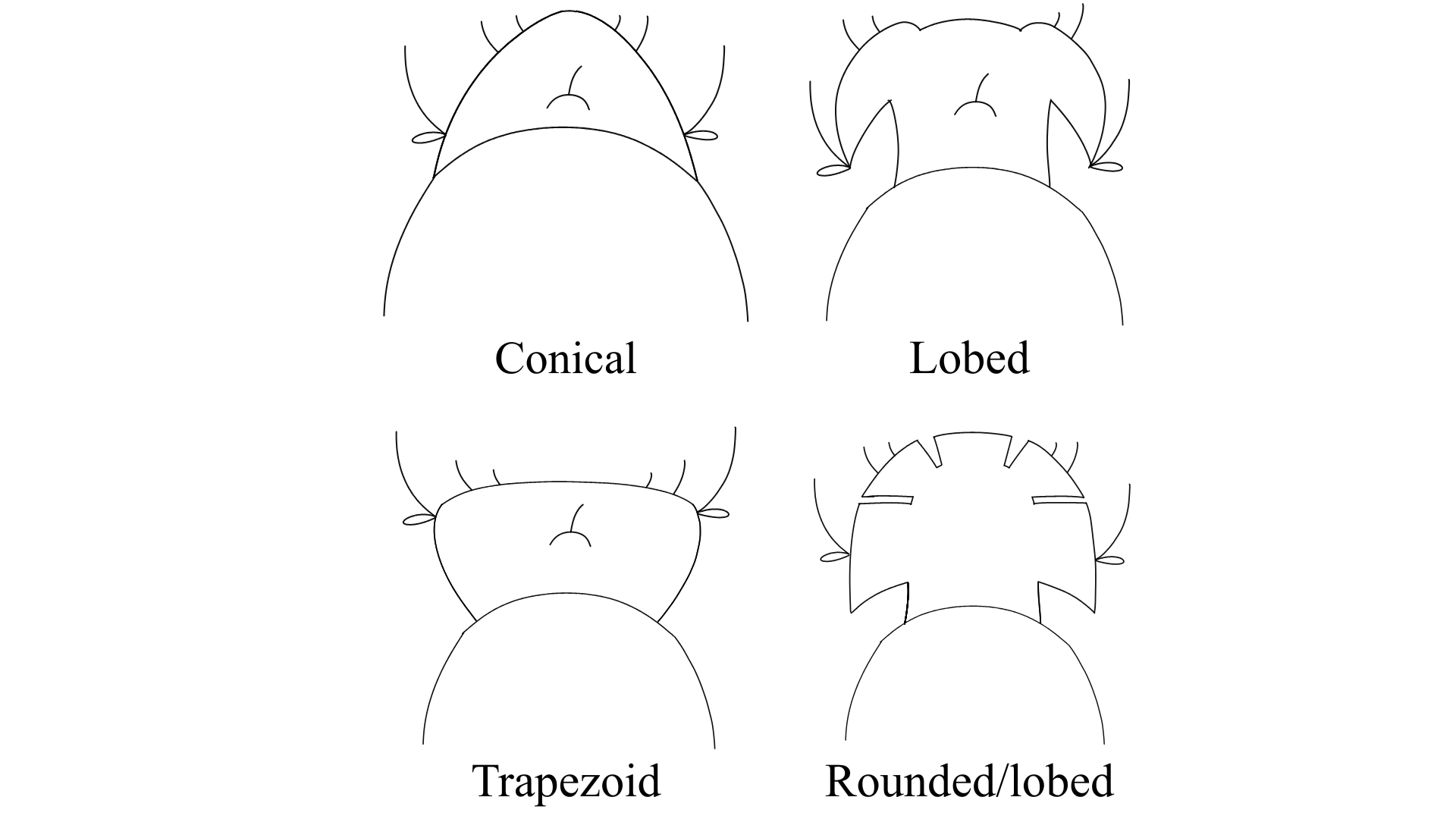
Head Structure

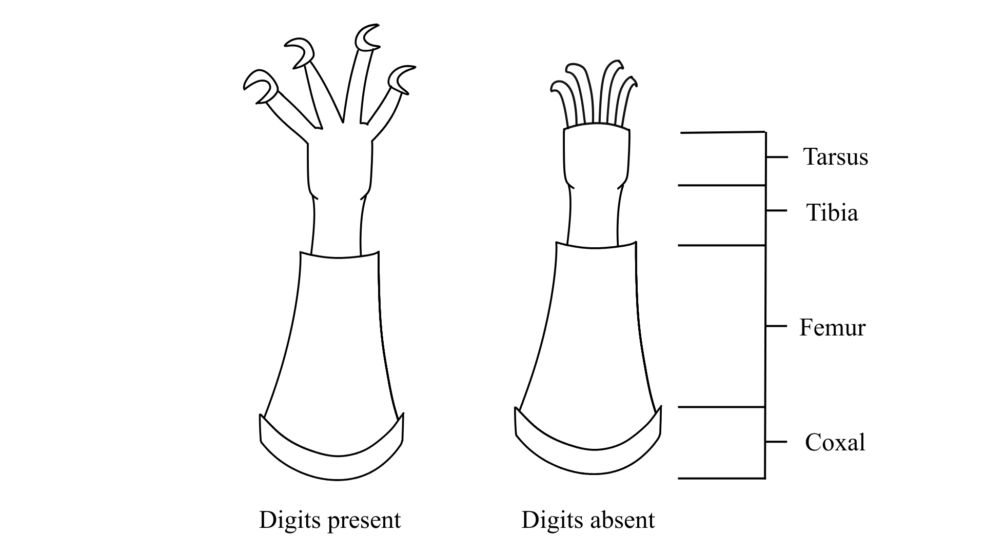
Digits

Note: If specimen has pads instead of digits, select “Pads”

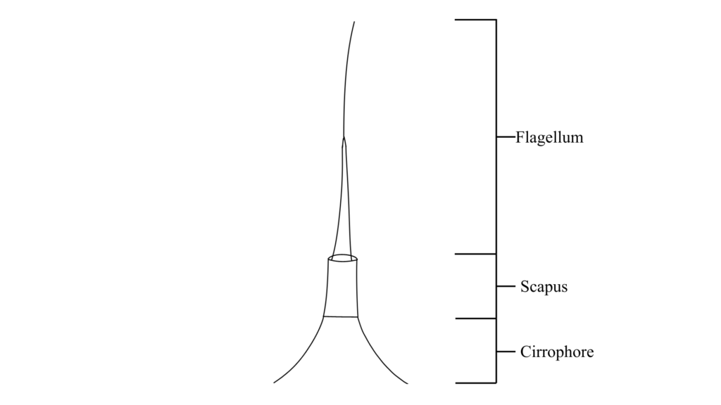
Cephalic cirri flagellum form

Modified image from Renaud-Mornant 1982 0708 Renaud-Mornant J. 1982. Species Diversity in Marine Tardigrada. Proceedings of the Third International Symposium on the Tardigrada. 149-178

Note: If unsure of form, select “Present”

Cephalic cirri scapus form

Modified image from Renaud-Mornant 1982 0708 Renaud-Mornant J. 1982. Species Diversity in Marine Tardigrada. Proceedings of the Third International Symposium on the Tardigrada. 149-178

Note: If unsure of form, select “Present” 

Cephalic cirri cirrophore form

Modified image from Renaud-Mornant 1982 0708 Renaud-Mornant J. 1982. Species Diversity in Marine Tardigrada. Proceedings of the Third International Symposium on the Tardigrada. 149-178

Note: If unsure of form, select “indistinct” for cirrophore

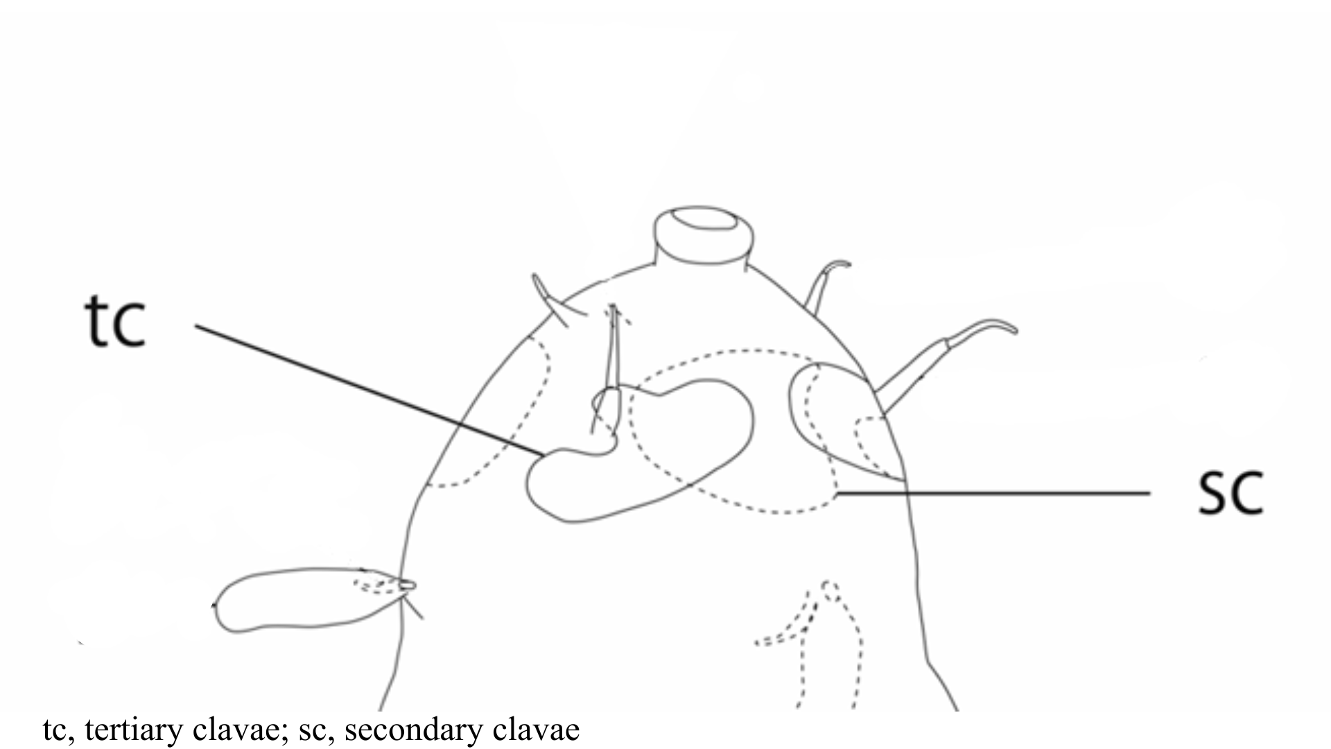
Secondary and tertiary clavae

Image from Fujimoto S, Hansen JG. 2019. Revision of Angursa (Arthrotardigrada: Styraconyxidae) with the description of a new species from Japan. European Journal of Taxonomy. 150: 1-19.

Note: Secondary clavae can have similar forms to primary clavae. However, secondary clavae can be flat structures of varying shapes around the mouth cone area

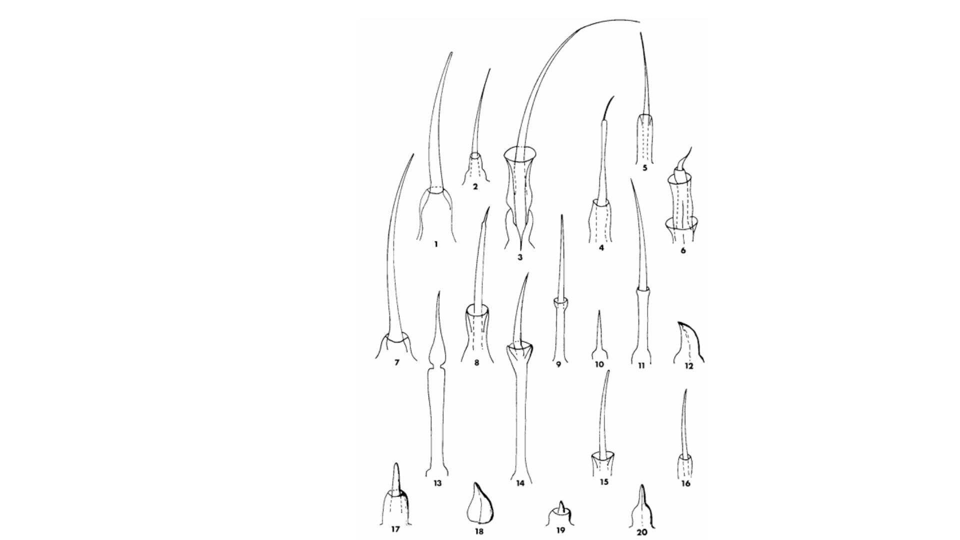
Generalized forms of primary clavae

Arthrotardigrada generalized primary clavae form

Note: Clavae forms can vary between species

Armoured plates?

Note: Select “Ventral plates” if visible.

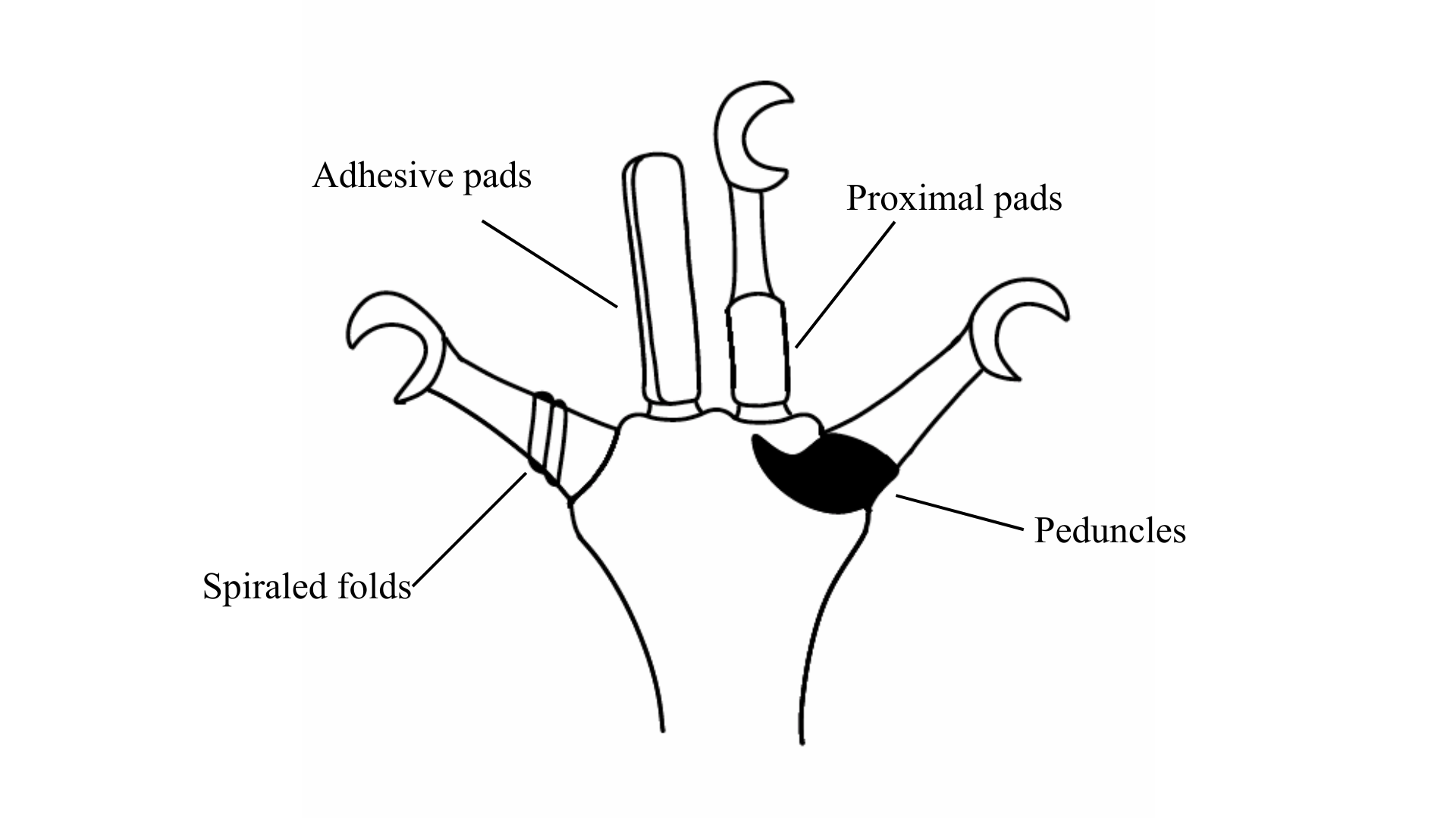
Additional digit accessory structures

Arthrotardigrada digit accessory structures

Note: Specimen can have some or none of these traits. Select those that apply.

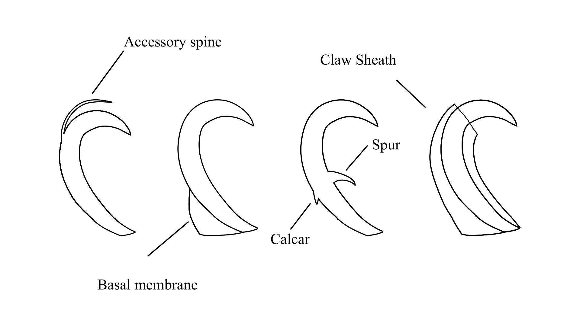
Additional claw accessory structures

Arthrotardigrada claw accessory structures

Note: Specimen can have some or none of these traits. Select those that apply.

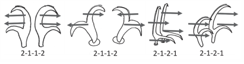
Claw Symmetry Relative To Median Plane Of Leg?

Image from Bingemer J, Hohberg K. 2017. An illustrated identification key to the eutardigrade species (Tardigrada, Eutardigrada) presently known from European soils. Soil Organisms. 89 (3): 127-149.

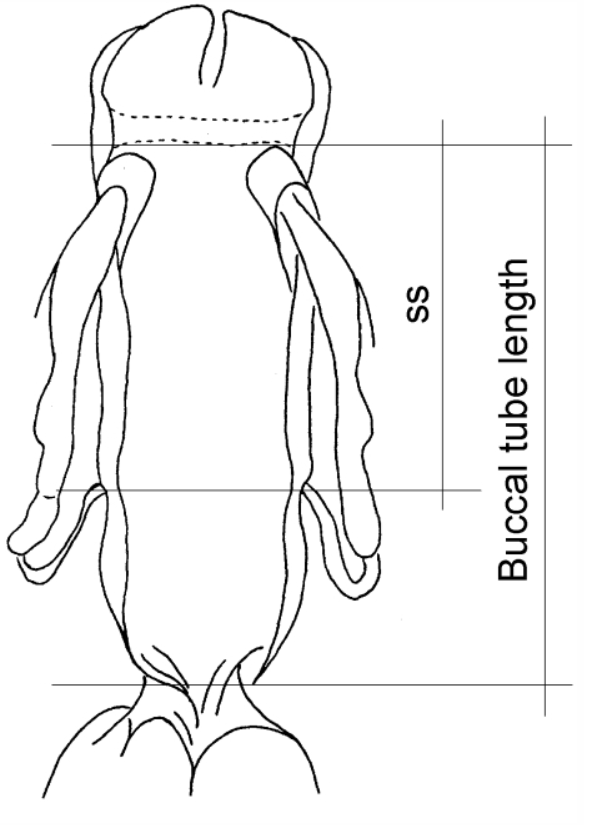
Stylet Support Insertion Point As Percentage Of Buccal Tube Length

Stylet support insertion point = ss divided by Buccal tube length, as %. Note anterior measurements begin at anterior margin of stylet sheaths, ss is centred where stylet supports reach buccal tube (this requires a good dorsal or lateral view for proper measurement)


Image from Tumanov DV. 2006. Five new species of the genus Milnesium (Tardigrada, Eutardigrada, Milnesiidae). Zootaxa. 1122: 1-23.

Buccal Tube Long, Mouth At Anterior Of A Protrusible Snout

Images from Pilato G, Binda MG. 2010. Definition of families, subfamilies, genera, and subgenera of the Eutardigrada, and keys to their identification. Zootaxa. 2404: 1-54.

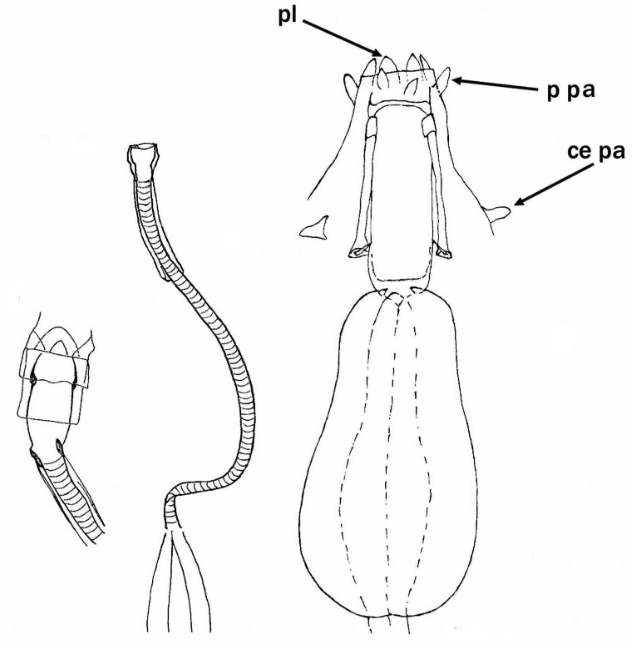
Pharyngeal Tube With Spiral Reinforcement

Images from Pilato G, Binda MG. 2010. Definition of families, subfamilies, genera, and subgenera of the Eutardigrada, and keys to their identification. Zootaxa. 2404: 1-54.

Peribuccal papillae?

Images from Pilato G, Binda MG. 2010. Definition of families, subfamilies, genera, and subgenera of the Eutardigrada, and keys to their identification. Zootaxa. 2404: 1-54.

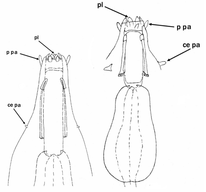
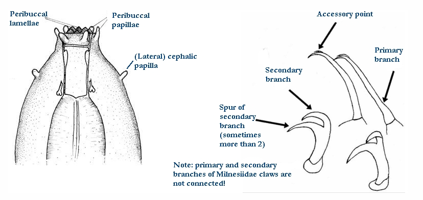
(Lateral) cephalic papillae, peribuccal papillae, Milnesiidae - type claws, exceptionally wide (and usually short) buccal tube?

Cephalic image modified from Nelson DR, Guidetti R, Rebecchi L. 2009. Tardigrada. Ch. 14 in Ecology and Classification of North American Freshwater Invertebrates, Thorp JH, Covich AP (eds.), 3rd ed.

Claw image modified from Pilato G, Binda MG. 2010. Definition of families, subfamilies, genera, and subgenera of the Eutardigrada, and keys to their identification. Zootaxa. 2404: 1-54.

Pharyngeal tube flexible?

Note: in a tardigrade with snout extended, the flex of the pharyngeal tube may not be obvious.


Images from Pilato G, Binda MG. 2010. Definition of families, subfamilies, genera, and subgenera of the Eutardigrada, and keys to their identification. Zootaxa. 2404: 1-54.

Serrated / dentate cuff / collar on legs IV

Left image from Kristensen RM. 1987. Generic revision of the Echiniscidae (Heterotardigrada), with a discussion of the origin of the family. pp. 261-335 in Bertolani R (ed). Biology of Tardigrades: Selected symposia and monographs.

Right Image from Richters F. 1926. Tardigrada. in Krumbach T. 1927. Handbuch der Zoologie, 3rd band, Walter de Gruyter & Co.

Leg sensory structures

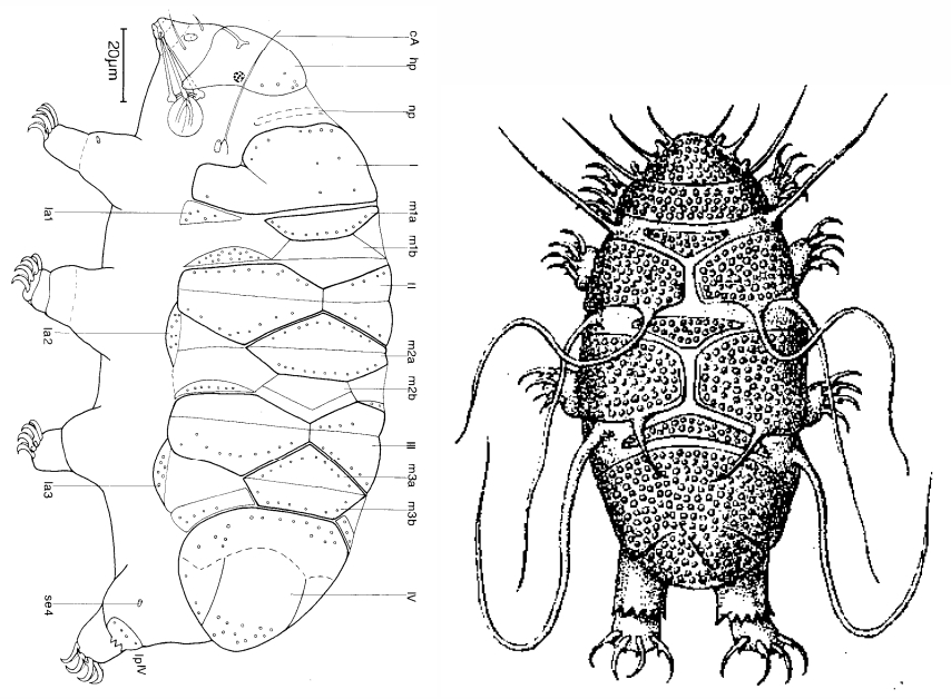
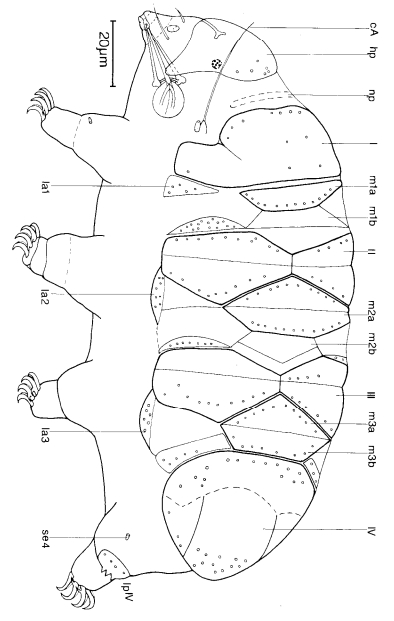
Typically papilla on leg IV (se4 in image) and variable shape on leg I


Image from Kristensen RM. 1987. Generic revision of the Echiniscidae (Heterotardigrada), with a discussion of the origin of the family. pp. 261-335 in Bertolani R (ed). Biology of Tardigrades: Selected symposia and monographs.

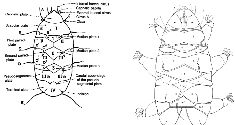
Trunk appendages

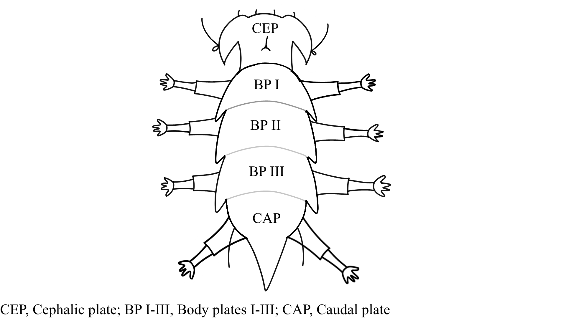
Appendages (after Cirrus A) labeled according to plate … B (Scapular plate), C (First segmental plate), D (Second segmental plate), E (terminal plate). “B” is lateral, “Bd” is dorsal

Be careful! There can be great variability within a population, with individuals (especially juveniles) lacking some appendages. When in doubt, focus on other characters first!


Left image from Ramazzotti G, Maucci W. 1983. Il phylum Tardigrada(III edizione riveduta e aggiornata). English translation by C. W. Beasley, 1995. Memorie dell’ Istituto Italiano di Idrobiologia 41: 1-1012.

Right image cropped from Kristensen RM. 1987. Generic revision of the Echiniscidae (Heterotardigrada), with a discussion of the origin of the family. pp. 261-335 in Bertolani R (ed). Biology of Tardigrades: Selected symposia and monographs.

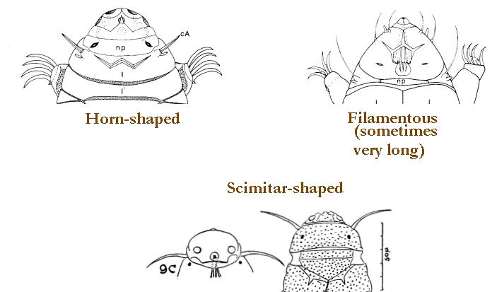
Cirrus A form

Images modified from Kristensen RM. 1987. Generic revision of the Echiniscidae (Heterotardigrada), with a discussion of the origin of the family. pp. 261-335 in Bertolani R (ed). Biology of Tardigrades: Selected symposia and monographs.

Lower image (Mopsechiniscus) modified from du Bois-Reymond Marcus E. 1944. Sobre tardigrados brasileiros. Communicaciones Zoologicas del Museo de Historia Natural de Montevideo. 1(13): 1-19 plus plates.

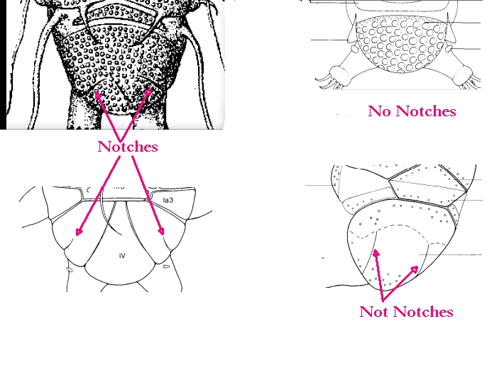
Terminal / caudal plate with two notches?

Images modified from Kristensen RM. 1987. Generic revision of the Echiniscidae (Heterotardigrada), with a discussion of the origin of the family. pp. 261-335 in Bertolani R (ed). Biology of Tardigrades: Selected symposia and monographs.

First image (Echiniscus) modified from Richters F. 1926. Tardigrada. in Krumbach T. 1927. Handbuch der Zoologie, 3rd band, Walter de Gruyter & Co.

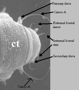
Inner & outer buccal cirri present

Image modified from Nelson DR, Guidetti R, Rebecchi L. 2009. Tardigrada. Ch. 14 in Ecology and Classification of North American Freshwater Invertebrates, Thorp JH, Covich AP (eds.), 3rd ed.

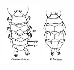
Pseudosegmental plate between last medial plate & terminal plate?

Note on 2nd image, what you see before the terminal plate is either no plate, or a full-width medial plate; don’t confuse the latter with a pseudosegmental plate!
Also, sometimes the pseudosegmental plate is paired, like segmental plates II & III. It’s still pseudosegmental!

Images from Clifford HF. 1991. Aquatic invertebrates of Alberta. University of Alberta Press, Alberta, Canada.

Segmental plates larger than intersegmental?

Image modified from Lindahl & Balser (1999), http://www.iwu.edu/~tardisdp/Keypage35.html

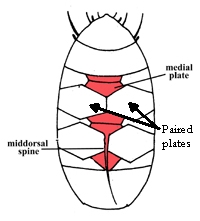
Any Paired Plates?

Modified from Lindahl K, Balser S.  1999. Key to tardigrade genera [Internet]. Available from: https://sun.iwu.edu/~tardisdp/Keypage33.html