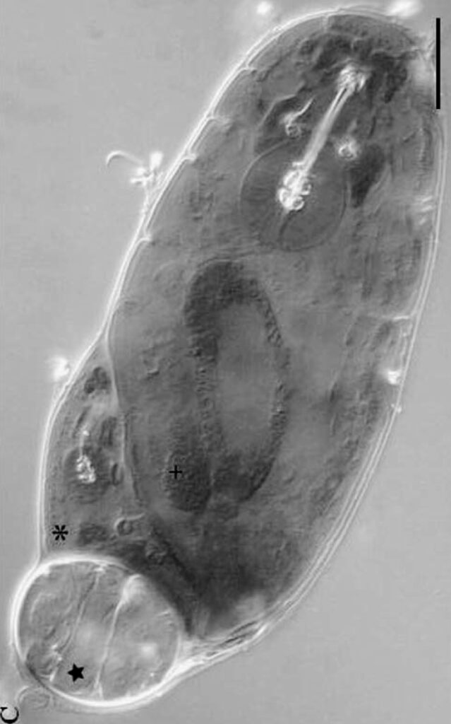
Cryobiotus

Dastych, H. (2019) Cryobiotus roswithae gen. n., sp. n., a new genus and species of glacier-dwelling tardigrades from northern Norway (Tardigrada, Panarthropoda). Entomologie heute, 31, 95–111.

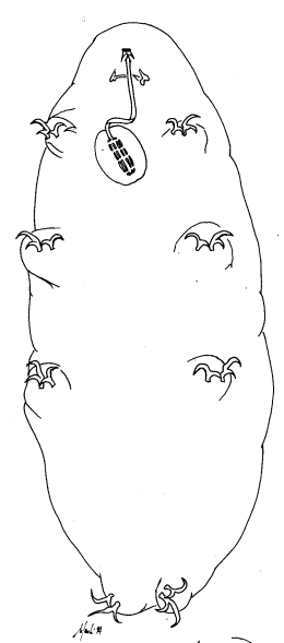
Kopakaius

Zawierucha, K., Stec, D., Dearden, P.K. & Shain, D.H. (2023) Two new tardigrade genera from New Zealand’s Southern Alp glaciers display morphological stasis and parallel evolution. Molecular Phylogenetics and Evolution 178, 107634. https://doi.org/10.1016/j.ympev.2022.107634

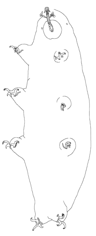
Kararehius

Zawierucha, K., Stec, D., Dearden, P.K. & Shain, D.H. (2023) Two new tardigrade genera from New Zealand’s Southern Alp glaciers display morphological stasis and parallel evolution. Molecular Phylogenetics and Evolution 178, 107634. https://doi.org/10.1016/j.ympev.2022.107634

Hypsibiinae

Hypsibiinae from Pilato 1969: “Unghie di tipo Isohypsibius o di tipo Hypsibius. Apparato boccale di tipo Hypsibius o di tipo Macrobiotus; bulbo faringeo provvisto di placoidi.” Translated: Claws of Isohypsibius or Hypsibius type. Buccal apparatus of Hypsibius or Macrobiotus type; pharyngeal bulb with placoids. Hypsibiinae from Bertolani et al. 2014: “Completely rigid buccal tube. Apophyses […]

Diphasconinae

“Eutardigrades with buccopharyngeal apparatus of Diphascon-type and anterior apophyses shaped as “blunt” or “semilunar” hooks (see Pilato 1987). Posteriodorsal apodeme (“drop-like” structure) present or absent, pharyngeal apophyses present. Asymmetrical claws of Hypsibiidae-type. Eggs either deposited into [shedded] cuticle and smooth or layed freely and with ornamented shells.” Dastych H. 1992. Paradiphascon manningi gen. n. sp. […]

Hypsibiidae

Hypsibiidae from Pilato 1969: “Senza alcuna appendice cefalica. Le due diplounghie di ciascuna zampa sono disposte asimmetricamente rispetto al piano mediano della zampa stessa e posssono essere di tipo Isohypsibius o di tipo Hypsibius. In ogni zampa le due diplounghie sono talvolta quasi uguali fra loro ma molto spesso profondamente diverse per forma e dimensioni. […]

Hypsibius

Genus description from Gąsiorek et al. 2018: “Six weakly outlined peribuccal lobes present. Apophyses for the insertion of stylet muscles in the shape of symmetrical hooks; and with well-developed caudal processes pointing diagonally (backwards and sideways […]). Pharyngeal apophyses and placoids present. Stylet furcae with the triangular base, thin arms and enlarged apices (sensu Pilato […]

Borealibius

Genus description from Pilato et al. 2006: “Claws of Hypsibius type; buccal–pharyngeal apparatus with rigid buccal tube without ventral lamina; in their caudal margins, the walls of the anterior portion of the buccal tube form well-developed transversal crests with very pronounced AISMs, almost cylindrical in shape with a large rounded or bilobed projection pointing caudally […]

Diphascon

Genus description from Plate 1888: “Diese Gattung hat grosse Aehnlichkeit mit Macrobiotus oberhäuseri, aber der kleine, runde Schlundkopf sitzt in der Mitte des Oesophagus. Das Gebiss ist zart. […] An jedem Bein 2 Doppelhaken, deren krallen am Grunde verwachsen sind.” Translated: This genus is very similar to Macrobiotus oberhäuseri, but the small, round pharynx sits […]

Mixibius

Genus description from Pilato 1992: “Hypsibiidae, organi ellittici sul cap assenti; unghie esterne di tipo Isohypsibius ed interne dello stesso tipo ma un po’modificate in quanto l’angolo fra unghia basale e ramo secondario è un po’maggiore di 90°; apparato bucco-faringeo con tubo boccale rigido, privo di sbarretta di rinforzo ventrale e con una lieve asimmetria rispetto al […]

Claw Symmetry Relative To Median Plane Of Leg?

Image from Bingemer J, Hohberg K. 2017. An illustrated identification key to the eutardigrade species (Tardigrada, Eutardigrada) presently known from European soils. Soil Organisms. 89 (3): 127-149.

Stylet Support Insertion Point As Percentage Of Buccal Tube Length

Stylet support insertion point = ss divided by Buccal tube length, as %. Note anterior measurements begin at anterior margin of stylet sheaths, ss is centred where stylet supports reach buccal tube (this requires a good dorsal or lateral view for proper measurement)


Image from Tumanov DV. 2006. Five new species of the genus Milnesium (Tardigrada, Eutardigrada, Milnesiidae). Zootaxa. 1122: 1-23.

Buccal Tube Long, Mouth At Anterior Of A Protrusible Snout

Images from Pilato G, Binda MG. 2010. Definition of families, subfamilies, genera, and subgenera of the Eutardigrada, and keys to their identification. Zootaxa. 2404: 1-54.

Pharyngeal Tube With Spiral Reinforcement

Images from Pilato G, Binda MG. 2010. Definition of families, subfamilies, genera, and subgenera of the Eutardigrada, and keys to their identification. Zootaxa. 2404: 1-54.

Peribuccal papillae?

Images from Pilato G, Binda MG. 2010. Definition of families, subfamilies, genera, and subgenera of the Eutardigrada, and keys to their identification. Zootaxa. 2404: 1-54.

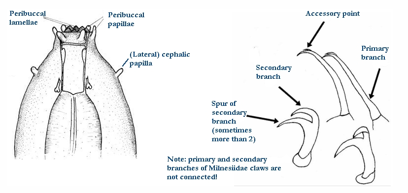
(Lateral) cephalic papillae, peribuccal papillae, Milnesiidae - type claws, exceptionally wide (and usually short) buccal tube?

Cephalic image modified from Nelson DR, Guidetti R, Rebecchi L. 2009. Tardigrada. Ch. 14 in Ecology and Classification of North American Freshwater Invertebrates, Thorp JH, Covich AP (eds.), 3rd ed.

Claw image modified from Pilato G, Binda MG. 2010. Definition of families, subfamilies, genera, and subgenera of the Eutardigrada, and keys to their identification. Zootaxa. 2404: 1-54.

Pharyngeal tube flexible?

Note: in a tardigrade with snout extended, the flex of the pharyngeal tube may not be obvious.


Images from Pilato G, Binda MG. 2010. Definition of families, subfamilies, genera, and subgenera of the Eutardigrada, and keys to their identification. Zootaxa. 2404: 1-54.

Serrated / dentate cuff / collar on legs IV

Left image from Kristensen RM. 1987. Generic revision of the Echiniscidae (Heterotardigrada), with a discussion of the origin of the family. pp. 261-335 in Bertolani R (ed). Biology of Tardigrades: Selected symposia and monographs.

Right Image from Richters F. 1926. Tardigrada. in Krumbach T. 1927. Handbuch der Zoologie, 3rd band, Walter de Gruyter & Co.

Leg sensory structures

Typically papilla on leg IV (se4 in image) and variable shape on leg I


Image from Kristensen RM. 1987. Generic revision of the Echiniscidae (Heterotardigrada), with a discussion of the origin of the family. pp. 261-335 in Bertolani R (ed). Biology of Tardigrades: Selected symposia and monographs.

Trunk appendages

Appendages (after Cirrus A) labeled according to plate … B (Scapular plate), C (First segmental plate), D (Second segmental plate), E (terminal plate). “B” is lateral, “Bd” is dorsal

Be careful! There can be great variability within a population, with individuals (especially juveniles) lacking some appendages. When in doubt, focus on other characters first!


Left image from Ramazzotti G, Maucci W. 1983. Il phylum Tardigrada(III edizione riveduta e aggiornata). English translation by C. W. Beasley, 1995. Memorie dell’ Istituto Italiano di Idrobiologia 41: 1-1012.

Right image cropped from Kristensen RM. 1987. Generic revision of the Echiniscidae (Heterotardigrada), with a discussion of the origin of the family. pp. 261-335 in Bertolani R (ed). Biology of Tardigrades: Selected symposia and monographs.

Cirrus A form

Images modified from Kristensen RM. 1987. Generic revision of the Echiniscidae (Heterotardigrada), with a discussion of the origin of the family. pp. 261-335 in Bertolani R (ed). Biology of Tardigrades: Selected symposia and monographs.

Lower image (Mopsechiniscus) modified from du Bois-Reymond Marcus E. 1944. Sobre tardigrados brasileiros. Communicaciones Zoologicas del Museo de Historia Natural de Montevideo. 1(13): 1-19 plus plates.

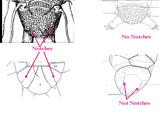
Terminal / caudal plate with two notches?

Images modified from Kristensen RM. 1987. Generic revision of the Echiniscidae (Heterotardigrada), with a discussion of the origin of the family. pp. 261-335 in Bertolani R (ed). Biology of Tardigrades: Selected symposia and monographs.

First image (Echiniscus) modified from Richters F. 1926. Tardigrada. in Krumbach T. 1927. Handbuch der Zoologie, 3rd band, Walter de Gruyter & Co.

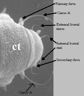
Inner & outer buccal cirri present

Image modified from Nelson DR, Guidetti R, Rebecchi L. 2009. Tardigrada. Ch. 14 in Ecology and Classification of North American Freshwater Invertebrates, Thorp JH, Covich AP (eds.), 3rd ed.

Pseudosegmental plate between last medial plate & terminal plate?

Note on 2nd image, what you see before the terminal plate is either no plate, or a full-width medial plate; don’t confuse the latter with a pseudosegmental plate!
Also, sometimes the pseudosegmental plate is paired, like segmental plates II & III. It’s still pseudosegmental!

Images from Clifford HF. 1991. Aquatic invertebrates of Alberta. University of Alberta Press, Alberta, Canada.

Segmental plates larger than intersegmental?

Image modified from Lindahl & Balser (1999), http://www.iwu.edu/~tardisdp/Keypage35.html

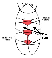
Any Paired Plates?

Modified from Lindahl K, Balser S.  1999. Key to tardigrade genera [Internet]. Available from: https://sun.iwu.edu/~tardisdp/Keypage33.html