Insuetifurca

Abe W. Occurrence of the semiterrestrial tardigrade Insuetifurca austronipponica (Eutardigrada: Macrobiotidae) on Izena Island, the Ryukyu Archipelago, southwestern Japan. Annual Report of Premedical Sciences, Dokkyo Medical University. 2: 43-46.

Class : Eutardigrada

Order : Macrobiotoidea

Family : Macrobiotidea

Genus : Insuetifurca

Macrobiotoidea from Marley et al. 2011: “Parachela; claw pairs symmetrical (2112); AISM generally asymmetrical, due to the ventral lamina, with lateral caudal processes of crests and hooks.”

Macrobiotoidea from Bertolani et al. 2014: “Double claws symmetrical with respect to the median plane of the leg (sequence 2112); double claw of each leg similar in shape and size; each double claw characterized by the presence of a peculiar stalk (peduncle) with cylindrical or laminar shape; 10 peribuccal lamellae or papulae; buccal tube strengthened by a ventral lamina. Eggs laid freely and always surrounded by an ornamented shell.”

Macrobiotidae from Thulin 1928: “Fam. Macrobiotidae zeichnet sich dadurch aus, dass Sinnesanhänge am Kopfe fehlen. Die Krallen sind zweihakig. Die hintere an jeder Extremität kann mehr oder weniger vollständig in Basal- und Terminal-kralle aufgeteilt sein, die vordere ist dagegen niemals in dieser Weise differenziert. Die Länge und Weite der Mundröhre variiert erheblich innerhalb dieser Familie, ebenso die Form des Schlundkopfes. Die Mundöffnung ist nicht durch Klappen verschliessbar, aber bisweilen (bei vielen Macrobiotus-Arten) von einem Lamellenkranz umgeben. Die Stilette sind mehr oder weniger stark bogenförmig gekrümmt, mit der Konvexität des Bogens medialwärts gewendet, und kräftiger als bei den übrigen Tardigraden.”

Translation: Fam. Macrobiotidae is characterized by the fact that sensory appendages are missing on the head. The claws are double-hooked. The posterior of each claw may be more or less completely divided into basal and terminal [secondary] claws, whereas the anterior [primary branch] is never differentiated in this way. The length and width of the mouth tube varies considerably within this family, as does the shape of the pharynx. The mouth opening is not closed by flaps, but sometimes (in many Macrobiotus species) surrounded by a lamellae. The stylets are more or less curved, with the convexity of the arch turned towards the medial direction, and stronger than in other tardigrades.

Macrobiotidae from Guidetti et al. 2005: “Parachela with double-claws of each leg similar in shape and size and symmetrical with respect to the median plane of the leg (sequence 2112). Y-shaped claws, with claw branches fused over a tract of variable length (common tract). Ten peribuccal structures (lamellae or papulae) present. Epicuticular layer compact and uniform, without pillar-like structures.”

Macrobiotidae from Bertolani et al. 2014: “Double claws Y-shaped, with the two branches forming an evident common basal tract of variable length. Buccal tube completely rigid, or caudally annulated.”

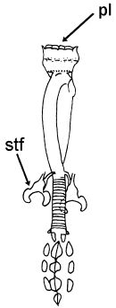
Genus description from Guidetti & Pilato 2003: “Diploclaws of a slightly modified ‘hufelandi type’ with the basal portion subdivided into a very short stalk and a large distal portion less sclerified than the common tract; lunules present; buccal-pharyngeal tube subdivided into an anterior rigid portion (Provided with a ventral strengthening bar) and a flexible caudal portion with a spiral cuticular thickening; peribuccal lamellae present; stylet furcae well developed and of a peculiar shape, with the posterior processes arched and converging backwards.”


Pilato G, Binda MG. 2010. Definition of families, subfamilies, genera, and subgenera of the Eutardigrada, and keys to their identification. Zootaxa. 2404: 1-54.

Citations:

Bertolani R, Guidetti R, Marchioro T, Altiero T, Rebecchi L, Cesari M. 2014. Phylogeny of Eutardigrada: New molecular data and their morphological support lead to the identification of new evolutionary lineages. Molecular Phylogenetics and Evolution. 76: 110-126.

Guidetti R, Gandolfi A, Rossi V, Bertolani R. 2005. Phylogenetic analysis of Macrobiotidae (Eutardigrada, Parachela): a combined morphological and molecular approach. Zoologica Scripta. 34 (3): 235-244.

Guidetti R, Pilato G. 2003. Revision of the genus Pseudodiphascon (Tardigrada, Macrobiotidae), with the erection of three new genera. Journal of Natural History. 37 (14): 1679-1690.

Marley NJ, McInnes SJ, Sands CJ. 2011. Phylum Tardigrada: A re-evaluation of the Parachela. Zootaxa. 2819: 51-64.

Thulin G. 1928. Über die Phylogenie und das System der Tardigraden. Hereditas. 11: 207–266. https://doi.org/10.1111/j.1601-5223.1928.tb02488.x

Claw Symmetry Relative To Median Plane Of Leg?

Image from Bingemer J, Hohberg K. 2017. An illustrated identification key to the eutardigrade species (Tardigrada, Eutardigrada) presently known from European soils. Soil Organisms. 89 (3): 127-149.

Stylet Support Insertion Point As Percentage Of Buccal Tube Length

Stylet support insertion point = ss divided by Buccal tube length, as %. Note anterior measurements begin at anterior margin of stylet sheaths, ss is centred where stylet supports reach buccal tube (this requires a good dorsal or lateral view for proper measurement)


Image from Tumanov DV. 2006. Five new species of the genus Milnesium (Tardigrada, Eutardigrada, Milnesiidae). Zootaxa. 1122: 1-23.

Buccal Tube Long, Mouth At Anterior Of A Protrusible Snout

Images from Pilato G, Binda MG. 2010. Definition of families, subfamilies, genera, and subgenera of the Eutardigrada, and keys to their identification. Zootaxa. 2404: 1-54.

Pharyngeal Tube With Spiral Reinforcement

Images from Pilato G, Binda MG. 2010. Definition of families, subfamilies, genera, and subgenera of the Eutardigrada, and keys to their identification. Zootaxa. 2404: 1-54.

Peribuccal papillae?

Note: in a tardigrade with snout extended, the flex of the pharyngeal tube may not be obvious.


Images from Pilato G, Binda MG. 2010. Definition of families, subfamilies, genera, and subgenera of the Eutardigrada, and keys to their identification. Zootaxa. 2404: 1-54.

(Lateral) cephalic papillae, peribuccal papillae, Milnesiidae - type claws, exceptionally wide (and usually short) buccal tube?

Cephalic image modified from Nelson DR, Guidetti R, Rebecchi L. 2009. Tardigrada. Ch. 14 in Ecology and Classification of North American Freshwater Invertebrates, Thorp JH, Covich AP (eds.), 3rd ed.

Claw image modified from Pilato G, Binda MG. 2010. Definition of families, subfamilies, genera, and subgenera of the Eutardigrada, and keys to their identification. Zootaxa. 2404: 1-54.

Pharyngeal tube flexible?

Note: in a tardigrade with snout extended, the flex of the pharyngeal tube may not be obvious.


Images from Pilato G, Binda MG. 2010. Definition of families, subfamilies, genera, and subgenera of the Eutardigrada, and keys to their identification. Zootaxa. 2404: 1-54.

Serrated / dentate cuff / collar on legs IV

Left image from Kristensen RM. 1987. Generic revision of the Echiniscidae (Heterotardigrada), with a discussion of the origin of the family. pp. 261-335 in Bertolani R (ed). Biology of Tardigrades: Selected symposia and monographs.

Right Image from Richters F. 1926. Tardigrada. in Krumbach T. 1927. Handbuch der Zoologie, 3rd band, Walter de Gruyter & Co.

Leg sensory structures

Typically papilla on leg IV (se4 in image) and variable shape on leg I


Image from Kristensen RM. 1987. Generic revision of the Echiniscidae (Heterotardigrada), with a discussion of the origin of the family. pp. 261-335 in Bertolani R (ed). Biology of Tardigrades: Selected symposia and monographs.

Trunk appendages

Appendages (after Cirrus A) labeled according to plate … B (Scapular plate), C (First segmental plate), D (Second segmental plate), E (terminal plate). “B” is lateral, “Bd” is dorsal

Be careful! There can be great variability within a population, with individuals (especially juveniles) lacking some appendages. When in doubt, choose “Unclear” to ignore this character!


Left image from Ramazzotti G, Maucci W. 1983. Il phylum Tardigrada(III edizione riveduta e aggiornata). English translation by C. W. Beasley, 1995. Memorie dell’ Istituto Italiano di Idrobiologia 41: 1-1012.

Right image cropped from Kristensen RM. 1987. Generic revision of the Echiniscidae (Heterotardigrada), with a discussion of the origin of the family. pp. 261-335 in Bertolani R (ed). Biology of Tardigrades: Selected symposia and monographs.

Cirrus A form

Images modified from Kristensen RM. 1987. Generic revision of the Echiniscidae (Heterotardigrada), with a discussion of the origin of the family. pp. 261-335 in Bertolani R (ed). Biology of Tardigrades: Selected symposia and monographs.

Lower image (Mopsechiniscus) modified from du Bois-Reymond Marcus E. 1944. Sobre tardigrados brasileiros. Communicaciones Zoologicas del Museo de Historia Natural de Montevideo. 1(13): 1-19 plus plates.

Terminal / caudal plate with two notches?

Images modified from Kristensen RM. 1987. Generic revision of the Echiniscidae (Heterotardigrada), with a discussion of the origin of the family. pp. 261-335 in Bertolani R (ed). Biology of Tardigrades: Selected symposia and monographs.

First image (Echiniscus) modified from Richters F. 1926. Tardigrada. in Krumbach T. 1927. Handbuch der Zoologie, 3rd band, Walter de Gruyter & Co.

Inner & outer buccal cirri present

Image modified from Nelson DR, Guidetti R, Rebecchi L. 2009. Tardigrada. Ch. 14 in Ecology and Classification of North American Freshwater Invertebrates, Thorp JH, Covich AP (eds.), 3rd ed.

Pseudosegmental plate between last medial plate & terminal plate?

Note on 2nd image, what you see before the terminal plate is either no plate, or a full-width medial plate; don’t confuse the latter with a pseudosegmental plate!
Also, sometimes the pseudosegmental plate is paired, like segmental plates II & III. It’s still pseudosegmental!

Images from Clifford HF. 1991. Aquatic invertebrates of Alberta. University of Alberta Press, Alberta, Canada.

Segmental plates larger than intersegmental?

Image modified from Lindahl & Balser (1999), http://www.iwu.edu/~tardisdp/Keypage35.html

Any Paired Plates?

Images from Kristensen RM. 1987. Generic revision of the Echiniscidae (Heterotardigrada), with a discussion of the origin of the family. pp. 261-335