Eohypsibius

Kristensen RM. 1982. New aberrant eutardigrades from homothermic springs on Disko Island, West Greenland. In Proceedings of the Third International Symposium on Tardigrada, Tennessee (pp. 203 - 220).

Class : Eutardigrade

Order : Eohypsibioidea

Family : Eohypsibiidae

Genus : Eohypsibius

Eohypsibioidea from Bertolani & Kristensen 1987 in Marley et al. 2011: “Parachela; claws asymmetric (2121); Eohypsibiidae-type claw pairs; apophysis for the insertion of the stylet muscles (AISM) ridged with lateral caudal processes of crests and hooks.”

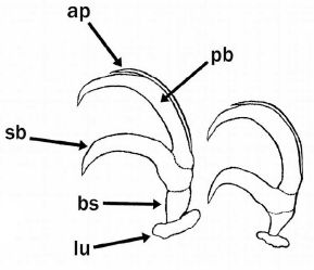
Eohypsibioidea from Bertolani et al. 2014: “Double claws of Eohypsibius type (i.e. subdivided into three distinct sections, one on top of the other), and asymmetrical with respect to the median plane of the leg (sequence 2121), with the ability to rotate on their base; double claws similar in size and shape on the same leg; 14 peribuccal lamellae; buccal tube completely rigid, or caudally annulated, in any case with dorsal and ventral crest-shaped apophyses for the insertion of the stylet muscles. Eggs laid freely and surrounded by an ornamented shell.”

Eohypsibiidae from Marley et al. 2011: “Eohypsibiidae-type claws, which are clearly delineated by septa, in linear order, from basal section, secondary branch and primary branch. The angle between basal section, secondary branch and primary branches are different between claws on the same leg and the internal claw can rotate on its base by 180°.”

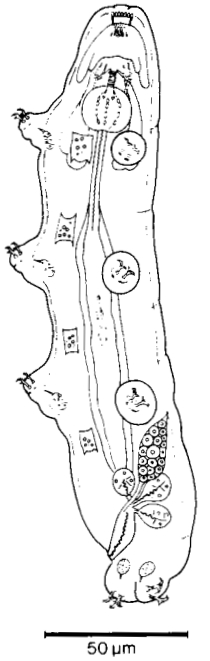
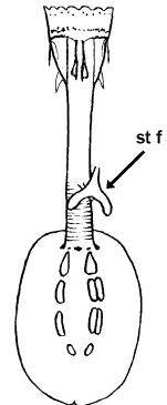
Genus description from Kristensen 1982: “An extremely slender calohypsibid with reduced legs and claws. Mouth opening large with 14-16 lamellae. A complete set of teeth in the buccal lumen; annulated buccal tube. Apophyses ventrally and dorsally hook-shaped and attached to the stylet retractor muscles. Claws of the Calohypsibius-type, but with lunules. Malpighian tubules ovoid with a small stalk. […] Discussion: The genus Eohypsibius has a buccal apparatus related to Pseudodiphascon (Christenberry & Higgins, 1979) and a claw system similar to Calohypsibius. The annulation of the buccal tube can only be seen after clearing the animals in polyvinyl lactophenol.”

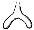
Additional genus notes from Pilato & Binda 1996: “…the furca of the stylets has an unusual shape. We noted that: a) the two branches are slightly arched; b) their apices are not clearly swollen.” Need modification by Trygvadóttir & Kristensen 2011.

Pilato G, Binda MG. 2010. Definition of families, subfamilies, genera, and subgenera of the Eutardigrada, and keys to their identification. Zootaxa. 2404: 1-54.

Citations:

Bertolani R, Guidetti R, Marchioro T, Altiero T, Rebecchi L, Cesari M. 2014. Phyloeny of Eutardigrada: New molecular data and their morphological support lead to the identification of new evolutionary lineages. Molecular Phylogenetics and Evolution. 76: 110-126.

Bertolani R, Kristensen RM. 1987. New records of Eohypsibius nadjae Kristensen, 1982, and revision of the taxonomic position of two genera of Eutardigrada (Tardigrada). In “Biology of Tardigrades.” (Ed. R Bertolani) pp. 359-372. (U.Z.I., I Mucchi: Modena, Italy).

Kristensen RM. 1982. New aberrant eutardigrades from homothermic springs on Disko Island,
West Greenland. In Proceedings of the Third International Symposium on Tardigrada, Tennessee (pp. 203 – 220).

Marley NJ, McInnes SJ, Sands CJ. 2011. Phylum Tardigrada: A re-evaluation of the Parachela. Zootaxa. 2819: 51-64.

Pilato G, Binda MG. 1996. Additional remarks to the description of some genera of eutardigrades. Bollettino delle Sedute della Accademia Gioenia di Scienze Naturali in Catania. 29 (351): 33-40.

Claw Symmetry Relative To Median Plane Of Leg?

Image from Bingemer J, Hohberg K. 2017. An illustrated identification key to the eutardigrade species (Tardigrada, Eutardigrada) presently known from European soils. Soil Organisms. 89 (3): 127-149.

Stylet Support Insertion Point As Percentage Of Buccal Tube Length

Stylet support insertion point = ss divided by Buccal tube length, as %. Note anterior measurements begin at anterior margin of stylet sheaths, ss is centred where stylet supports reach buccal tube (this requires a good dorsal or lateral view for proper measurement)


Image from Tumanov DV. 2006. Five new species of the genus Milnesium (Tardigrada, Eutardigrada, Milnesiidae). Zootaxa. 1122: 1-23.

Buccal Tube Long, Mouth At Anterior Of A Protrusible Snout

Images from Pilato G, Binda MG. 2010. Definition of families, subfamilies, genera, and subgenera of the Eutardigrada, and keys to their identification. Zootaxa. 2404: 1-54.

Pharyngeal Tube With Spiral Reinforcement

Images from Pilato G, Binda MG. 2010. Definition of families, subfamilies, genera, and subgenera of the Eutardigrada, and keys to their identification. Zootaxa. 2404: 1-54.

Peribuccal papillae?

Note: in a tardigrade with snout extended, the flex of the pharyngeal tube may not be obvious.


Images from Pilato G, Binda MG. 2010. Definition of families, subfamilies, genera, and subgenera of the Eutardigrada, and keys to their identification. Zootaxa. 2404: 1-54.

(Lateral) cephalic papillae, peribuccal papillae, Milnesiidae - type claws, exceptionally wide (and usually short) buccal tube?

Cephalic image modified from Nelson DR, Guidetti R, Rebecchi L. 2009. Tardigrada. Ch. 14 in Ecology and Classification of North American Freshwater Invertebrates, Thorp JH, Covich AP (eds.), 3rd ed.

Claw image modified from Pilato G, Binda MG. 2010. Definition of families, subfamilies, genera, and subgenera of the Eutardigrada, and keys to their identification. Zootaxa. 2404: 1-54.

Pharyngeal tube flexible?

Note: in a tardigrade with snout extended, the flex of the pharyngeal tube may not be obvious.


Images from Pilato G, Binda MG. 2010. Definition of families, subfamilies, genera, and subgenera of the Eutardigrada, and keys to their identification. Zootaxa. 2404: 1-54.

Serrated / dentate cuff / collar on legs IV

Left image from Kristensen RM. 1987. Generic revision of the Echiniscidae (Heterotardigrada), with a discussion of the origin of the family. pp. 261-335 in Bertolani R (ed). Biology of Tardigrades: Selected symposia and monographs.

Right Image from Richters F. 1926. Tardigrada. in Krumbach T. 1927. Handbuch der Zoologie, 3rd band, Walter de Gruyter & Co.

Leg sensory structures

Typically papilla on leg IV (se4 in image) and variable shape on leg I


Image from Kristensen RM. 1987. Generic revision of the Echiniscidae (Heterotardigrada), with a discussion of the origin of the family. pp. 261-335 in Bertolani R (ed). Biology of Tardigrades: Selected symposia and monographs.

Trunk appendages

Appendages (after Cirrus A) labeled according to plate … B (Scapular plate), C (First segmental plate), D (Second segmental plate), E (terminal plate). “B” is lateral, “Bd” is dorsal

Be careful! There can be great variability within a population, with individuals (especially juveniles) lacking some appendages. When in doubt, choose “Unclear” to ignore this character!


Left image from Ramazzotti G, Maucci W. 1983. Il phylum Tardigrada(III edizione riveduta e aggiornata). English translation by C. W. Beasley, 1995. Memorie dell’ Istituto Italiano di Idrobiologia 41: 1-1012.

Right image cropped from Kristensen RM. 1987. Generic revision of the Echiniscidae (Heterotardigrada), with a discussion of the origin of the family. pp. 261-335 in Bertolani R (ed). Biology of Tardigrades: Selected symposia and monographs.

Cirrus A form

Images modified from Kristensen RM. 1987. Generic revision of the Echiniscidae (Heterotardigrada), with a discussion of the origin of the family. pp. 261-335 in Bertolani R (ed). Biology of Tardigrades: Selected symposia and monographs.

Lower image (Mopsechiniscus) modified from du Bois-Reymond Marcus E. 1944. Sobre tardigrados brasileiros. Communicaciones Zoologicas del Museo de Historia Natural de Montevideo. 1(13): 1-19 plus plates.

Terminal / caudal plate with two notches?

Images modified from Kristensen RM. 1987. Generic revision of the Echiniscidae (Heterotardigrada), with a discussion of the origin of the family. pp. 261-335 in Bertolani R (ed). Biology of Tardigrades: Selected symposia and monographs.

First image (Echiniscus) modified from Richters F. 1926. Tardigrada. in Krumbach T. 1927. Handbuch der Zoologie, 3rd band, Walter de Gruyter & Co.

Inner & outer buccal cirri present

Image modified from Nelson DR, Guidetti R, Rebecchi L. 2009. Tardigrada. Ch. 14 in Ecology and Classification of North American Freshwater Invertebrates, Thorp JH, Covich AP (eds.), 3rd ed.

Pseudosegmental plate between last medial plate & terminal plate?

Note on 2nd image, what you see before the terminal plate is either no plate, or a full-width medial plate; don’t confuse the latter with a pseudosegmental plate!
Also, sometimes the pseudosegmental plate is paired, like segmental plates II & III. It’s still pseudosegmental!

Images from Clifford HF. 1991. Aquatic invertebrates of Alberta. University of Alberta Press, Alberta, Canada.

Segmental plates larger than intersegmental?

Image modified from Lindahl & Balser (1999), http://www.iwu.edu/~tardisdp/Keypage35.html

Any Paired Plates?

Images from Kristensen RM. 1987. Generic revision of the Echiniscidae (Heterotardigrada), with a discussion of the origin of the family. pp. 261-335