Antechiniscus

Kristensen RM. 1987. Generic revision of the Echiniscidae (Heterotardigrada), with a discussion of the origin of the family. pp. 261-335 in Bertolani R (ed). Biology of Tardigrades: Selected symposia and monographs.

Class : Heterotardigrada

Order : Echiniscoidae

Family : Echiniscidae

Genus : Antechiniscus

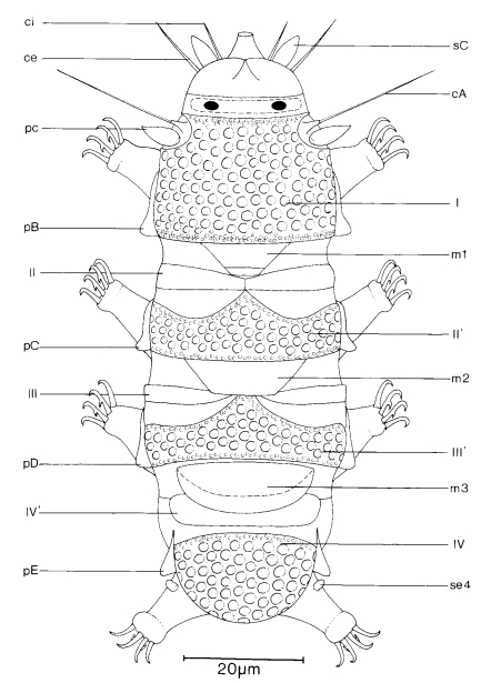
Antechiniscus, from Kristensen 1987: Echiniscidae with black eyes, unflexible buccal tube. Unpaired pseudosegmental plates (II’ and III’) present posterior to paired segmental plates II and III. Anterior to the caudal plate (IV) is a small bar-shaped pseudosegmental plate (IV’) which is slightly overlapped by a large undivided median plate (m 3). colour grey to light red; male small with enlarged primary and secondary clavae. … Length range – female: 140-197 µm; male: 80-123 µm. (based on Ramazzotti 1964): separated from Pseudechiniscus, because of the presence of the lateral structure, a straight line and a triangular papilla on each segment … most characterstic is the existence of additional pseudosegmental plates, and the very large median plate 3 with an internal suture.

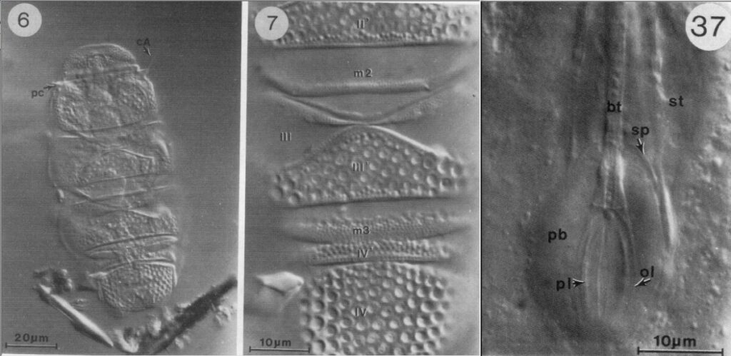
“Description: The ventral plates are absent. The head plate has lateral facets and medium coarse granulated cuticle. A small neck plate with a finely punctuated cuticle is distinctive. A large undivided scapular plate (I) has a thick and coarse granulated cuticle. The paired segmental plates (II and III) are thin with finely punctuated cuticle. The pseudosegmental elements (II’and III’) of the plates II and III are unpaired, strongly sclerotized and thick. The sculpture is the same as that found on the scapular plate and the posterior part of the caudal plate. The caudal plate (IV) divided into an anterior part with fine sculpture and a posterior part with a coarse granulated cuticle and lateral indentations (or not- ches). The pseudosegmental plate (IV) small and hidden by the overlapping me- dian plate 3. Both plates have the same granulation as the head plate. All three median plates are present. Median plate 1 undivided, but folded near the posterior margin. Very thin lateral intersegmental plates present in connection with median plate 1. Median plate 2 is triangular. Both plate 1 and 2 are finely sculptured. Median plate 3 is very large and has a round posterior margin which overlaps the pseudosegmental plate IV’.

The cephalic appendages (sense organs) are typical for the family Echiniscidae, but the male has enlarged primary clavae as in the family Oreellidae. The cirri A and primary clavae are located laterally between the anterior margin of the scapular plate (I) and the neck plate (np). The cirrus A has a distinct cirrophore and a medium sized flagellum. Each clava is inserted very close to the cirrophore of cirri A. The female has a short ovoid primary clava, the male has a longer primary clava with pointed tip. The secondary clavae are located ventrally and inserted close to the relatively long external cirri. The female has slender papillate secondary clavae as contrasted to the enlarged ovoid secondary clavae of the male. The internal cirri are short; both the external and internal cirri have a filiform flagellum with indistinct cirrophore. Trunk cirri can be absent, if so four pair of lateral triangular papillae are present. The papillae are connected by a bar- like structure and are homologous with cirri B, C, D and E found in other members of Echiniscidae. A pillow shaped structure is located lateroventrally between the pseudosegmental plates IV’ and the caudal plates. The only sense organ on the leg is a small papilla on leg IV. Only the internal claws have a secondary spur.

The buccal apparatus has a tube, which extends about half the distance in- to the pharyngeal bulb. The outer walls of the tube bends slightly within the bulb, so the distance between the outer and inner wall of the tube is thickest near the terminal end. The inner wall is continuous with the bar-shaped placoids, the outer wall remains as a thin cuticular lining. The buccal tube is inflexible, the stylets are long and inserted on the pharyngeal tube. The stylet supports are tiny, without CaCO3, and are located very close to the anterior margin of the pharyngeal bulb. Males are present in the two species of Antechiniscus from South America. They are very tiny, about half the size of females. Male gonopore is a small papilla with a crescent opening, testis is a relatively large sac with rod-headed sperms.”

(Similar to Pseudechiniscus, but additional plates.)

Kristensen RM. 1987. Generic revision of the Echiniscidae (Heterotardigrada), with a discussion of the origin of the family. pp. 261-335 in Bertolani R (ed). Biology of Tardigrades: Selected symposia and monographs.

Citations:

Kristensen RM. 1987. Generic revision of the Echiniscidae (Heterotardigrada), with a discussion of the origin of the family. pp. 261-335 in Bertolani R (ed). Biology of Tardigrades: Selected symposia and monographs.

Claw Symmetry Relative To Median Plane Of Leg?

Image from Bingemer J, Hohberg K. 2017. An illustrated identification key to the eutardigrade species (Tardigrada, Eutardigrada) presently known from European soils. Soil Organisms. 89 (3): 127-149.

Stylet Support Insertion Point As Percentage Of Buccal Tube Length

Stylet support insertion point = ss divided by Buccal tube length, as %. Note anterior measurements begin at anterior margin of stylet sheaths, ss is centred where stylet supports reach buccal tube (this requires a good dorsal or lateral view for proper measurement)


Image from Tumanov DV. 2006. Five new species of the genus Milnesium (Tardigrada, Eutardigrada, Milnesiidae). Zootaxa. 1122: 1-23.

Buccal Tube Long, Mouth At Anterior Of A Protrusible Snout

Images from Pilato G, Binda MG. 2010. Definition of families, subfamilies, genera, and subgenera of the Eutardigrada, and keys to their identification. Zootaxa. 2404: 1-54.

Pharyngeal Tube With Spiral Reinforcement

Images from Pilato G, Binda MG. 2010. Definition of families, subfamilies, genera, and subgenera of the Eutardigrada, and keys to their identification. Zootaxa. 2404: 1-54.

Peribuccal papillae?

Note: in a tardigrade with snout extended, the flex of the pharyngeal tube may not be obvious.


Images from Pilato G, Binda MG. 2010. Definition of families, subfamilies, genera, and subgenera of the Eutardigrada, and keys to their identification. Zootaxa. 2404: 1-54.

(Lateral) cephalic papillae, peribuccal papillae, Milnesiidae - type claws, exceptionally wide (and usually short) buccal tube?

Cephalic image modified from Nelson DR, Guidetti R, Rebecchi L. 2009. Tardigrada. Ch. 14 in Ecology and Classification of North American Freshwater Invertebrates, Thorp JH, Covich AP (eds.), 3rd ed.

Claw image modified from Pilato G, Binda MG. 2010. Definition of families, subfamilies, genera, and subgenera of the Eutardigrada, and keys to their identification. Zootaxa. 2404: 1-54.

Pharyngeal tube flexible?

Note: in a tardigrade with snout extended, the flex of the pharyngeal tube may not be obvious.


Images from Pilato G, Binda MG. 2010. Definition of families, subfamilies, genera, and subgenera of the Eutardigrada, and keys to their identification. Zootaxa. 2404: 1-54.

Serrated / dentate cuff / collar on legs IV

Left image from Kristensen RM. 1987. Generic revision of the Echiniscidae (Heterotardigrada), with a discussion of the origin of the family. pp. 261-335 in Bertolani R (ed). Biology of Tardigrades: Selected symposia and monographs.

Right Image from Richters F. 1926. Tardigrada. in Krumbach T. 1927. Handbuch der Zoologie, 3rd band, Walter de Gruyter & Co.

Leg sensory structures

Typically papilla on leg IV (se4 in image) and variable shape on leg I


Image from Kristensen RM. 1987. Generic revision of the Echiniscidae (Heterotardigrada), with a discussion of the origin of the family. pp. 261-335 in Bertolani R (ed). Biology of Tardigrades: Selected symposia and monographs.

Trunk appendages

Appendages (after Cirrus A) labeled according to plate … B (Scapular plate), C (First segmental plate), D (Second segmental plate), E (terminal plate). “B” is lateral, “Bd” is dorsal

Be careful! There can be great variability within a population, with individuals (especially juveniles) lacking some appendages. When in doubt, choose “Unclear” to ignore this character!


Left image from Ramazzotti G, Maucci W. 1983. Il phylum Tardigrada(III edizione riveduta e aggiornata). English translation by C. W. Beasley, 1995. Memorie dell’ Istituto Italiano di Idrobiologia 41: 1-1012.

Right image cropped from Kristensen RM. 1987. Generic revision of the Echiniscidae (Heterotardigrada), with a discussion of the origin of the family. pp. 261-335 in Bertolani R (ed). Biology of Tardigrades: Selected symposia and monographs.

Cirrus A form

Images modified from Kristensen RM. 1987. Generic revision of the Echiniscidae (Heterotardigrada), with a discussion of the origin of the family. pp. 261-335 in Bertolani R (ed). Biology of Tardigrades: Selected symposia and monographs.

Lower image (Mopsechiniscus) modified from du Bois-Reymond Marcus E. 1944. Sobre tardigrados brasileiros. Communicaciones Zoologicas del Museo de Historia Natural de Montevideo. 1(13): 1-19 plus plates.

Terminal / caudal plate with two notches?

Images modified from Kristensen RM. 1987. Generic revision of the Echiniscidae (Heterotardigrada), with a discussion of the origin of the family. pp. 261-335 in Bertolani R (ed). Biology of Tardigrades: Selected symposia and monographs.

First image (Echiniscus) modified from Richters F. 1926. Tardigrada. in Krumbach T. 1927. Handbuch der Zoologie, 3rd band, Walter de Gruyter & Co.

Inner & outer buccal cirri present

Image modified from Nelson DR, Guidetti R, Rebecchi L. 2009. Tardigrada. Ch. 14 in Ecology and Classification of North American Freshwater Invertebrates, Thorp JH, Covich AP (eds.), 3rd ed.

Pseudosegmental plate between last medial plate & terminal plate?

Note on 2nd image, what you see before the terminal plate is either no plate, or a full-width medial plate; don’t confuse the latter with a pseudosegmental plate!
Also, sometimes the pseudosegmental plate is paired, like segmental plates II & III. It’s still pseudosegmental!

Images from Clifford HF. 1991. Aquatic invertebrates of Alberta. University of Alberta Press, Alberta, Canada.

Segmental plates larger than intersegmental?

Image modified from Lindahl & Balser (1999), http://www.iwu.edu/~tardisdp/Keypage35.html

Any Paired Plates?

Images from Kristensen RM. 1987. Generic revision of the Echiniscidae (Heterotardigrada), with a discussion of the origin of the family. pp. 261-335