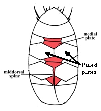
Isohypsibioidea from Marley et al. 2011: “Parachela. Claws asymmetrical (2121); Isohypsibius-type claw pairs; AISM ridged.”
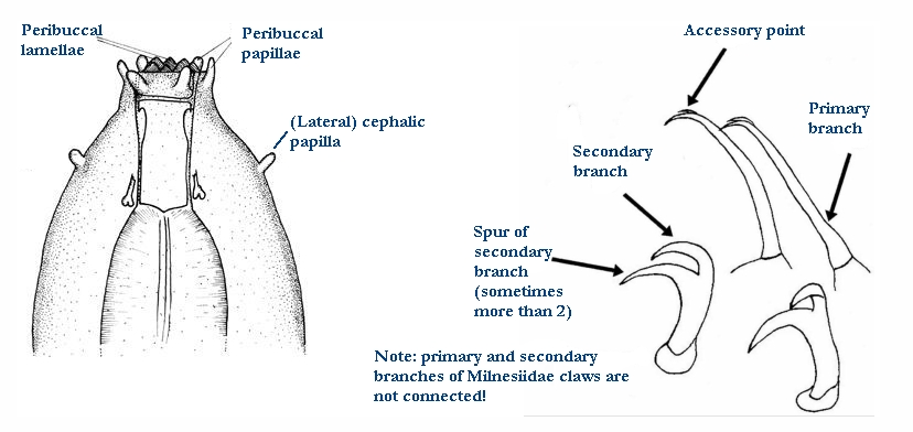
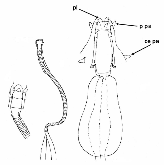
Isohypsibioidea from Bertolani et al. 2014: “Double claws asymmetrical with respect to the median plane of the leg (2121), normally with similar shape and size on each leg; double claws of the Isohypsibius type (secondary branch of the external claw inserted perpendicularly on the claw basal tract), or reduced from it: Hexapodibius type (very short, without common basal tract, with a base as large as the sum of the primary and secondary branch widths, and with an evident suture between primary and secondary branch); Haplomacrobiotus type (one branch only); completely absent (Apodibius). Buccal tube completely rigid (apart Paradiphascon; see below) and often relatively large, without (Dastychius, Eremobiotus, Halobiotus, Isohypsibius, Paradiphascon, Pseudobiotus, Thulinius) or with (Apodibius, Doryphoribius, Haplomacrobiotus, Haplohexapodibius, Hexapodibius, Parhexapodibius) ventral lamina. Eggs with smooth shell laid within the exuvium.”
Isohypsibiidae from Marley et al. 2011: “Isohypsibioidea. Claw pairs of similar size and shape. External and internal claws exhibiting articulation (the basal section and secondary branch form a solid unit while the primary branch and secondary branch articulate). Claws Isohypsibius-type, forming a right-angle between basal section and secondary branch. AISM ridge-like.”
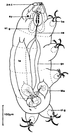
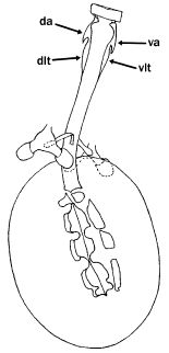
Genus description from Kristensen 1982: “Marine eutardigrades belonging to the family Hypsibiidae Pilato, 1969. The buccal tube has both a ventral list and a dorsal hook into which the stylet protractor muscles are inserted. The two double claws on each leg are heteronych. The external double claws with lunulae. The lunula of the internal claw of 1st-3rd pair of legs is transformaed into a cuticular barlike structure. The three Malpighian tubules are abnormally large.”
Note: Marine species only!
Genus additional notes from Pilato & Binda 1996: “…both the dorsal and ventral apophyses for the insertion of the stylet muscles are in shape of ‘semilunar hook’ with acute caudal apex. We noted also that both the dorsal and the ventral apophyses for the insertion of the stylet muscles are followed by a median longitudinal thickening. We cannot define the ventral thickening as a ‘ventral lamina’ identical to that of Macrobiotus. The bucco-pharyngeal apparatus is symmetrical with respect to the frontal plane.”

Citations:
Bertolani R, Guidetti R, Marchioro T, Altiero T, Rebecchi L, Cesari M. 2014. Phyloeny of Eutardigrada: New molecular data and their morphological support lead to the identification of new evolutionary lineages. Molecular Phylogenetics and Evolution. 76: 110-126.
Kristensen RM. 1982. The first record of cyclomorphosis in Tardigrada based on a new genus and species from Arctic meiobenthos. Journal of Zoological Systematics and Evolutionary Research 20(4):249 – 270.
Marley NJ, McInnes SJ, Sands CJ. 2011. Phylum Tardigrada: A re-evaluation of the Parachela. Zootaxa. 2819: 51-64.
Pilato G, Binda MG. 1996. Additional remarks to the description of some genera of eutardigrades. Bollettino delle Sedute della Accademia Gioenia di Scienze Naturali in Catania. 29 (351): 33-40.
