Hypechiniscus

Kristensen RM. 1987. Generic revision of the Echiniscidae (Heterotardigrada), with a discussion of the origin of the family. pp. 261-335 in Bertolani R (ed). Biology of Tardigrades: Selected symposia and monographs.

Order: Echiniscoidea

Family: Echiniscidae

Click taxa for descriptions

From Thulin 1928: “Echiniscidae with armor containing five median plates. End plate adjacent to the last median plate. Terminal notches present. Echiniscus gladiator MURRAY var. exarmatus MURR leads to this genus. The exarmatus form (from the Faroe Islands) that I have examined differs in some aspects from MURRAYS description: The animals are colorless with black eyes. The conjunctiva of the shell shows the same fine granulation as the plates. The end plate is faceted, with two lateral caudal facets and one unusually far reaching; the terminal notches are relatively short and wide. On the former deviations should no great weight be placed. As far as the end plate is concerned, MURRAY’s figure seems to me to say that he did not keep the terminal notches and the edges of the caudal facet apart, and that the latter caused him to regard the plate as being ‘extremely trifoliate’.”

Untranslated: “Echinisciden mit einem Plattenpanzer, der funf freie Schaltplatten enthalt. Endplatte an die letzte Schaltplatte grenzend. Kleeblattkerben vorhanden. Zu dieser Gattung führe ich Echiniscus gladiator MURRAY mit der var. exarmatus MURR. Die von mir untersuchte exarmatus-Form (von den Färöern stammend) weicht in einigen Punkten von MURRAYS Beschreibung ab: Die Tierchen sind farblos mit schwarzen Augen. Die Bindehaut des Panzers zeigt dieselbe feine Granulation wie die Platten. Die Endplatte ist fazettiert, mit zwei lateralen und einer sich ungewöhnlich weit hinauf erstreckenden kaudalen Fazette; die Kleeblattkerben sind verhältnismässig kurz und breit. Auf die ersteren Abweichungen dürfte kein grösseres Gewicht zu legen sein. Was die Endplatte betrifft, scheint mir MURRAYS Figur dafür zu sprechen, dass er die Kleeblattkerben und die Kanten der kaudalen Fazette nicht auseinander gehalten hat und dass die letzteren ihn veranlasst haben, die Platte als besonders ‘deeply trifoliate’ anzusehen.”

From Ramazzotti & Maucci: “There are visible inclusively 5 median plates; there exists the notches on the terminal plate.”

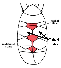
From Kristensen 1987: “Uncoloured Echiniscidae with ovoid black eyes; rigid buccal tube with a cuticular bulb anterior to a small pharyngeal bulb, stylet supports and ventral plates absent. Head plate large with a suture; scapular plates may be paired; paired segmental plates II and III, caudal plate divided in three pieces by sutures; all three median plates undivided. Both primary and secondary clavae papillate. Emended description: The dorsal cuticular plates indistinct and the sculpture is finely punctuated as the ventral cuticle. The suture of the large head plate nearly separates the plate into five pieces. A small neck plate is present. Lateral intersegmental plates are found between the segmental plates and median plates 1, 2 and 3. The cephalic cirri all have a large basis (cirrophore) from which a thinner filiform flagellum arise. The male has a very large secondary papillate clava, the primary clava insert very close to the cirrus A. Hypechiniscus gladiator has only a small tiny papilla located on the leg IV; H. papillifer has papilla on all four pairs of legs. The leg plates and collar are absent. Extraordinary is the location of the trunk appendage, when present it is located middorsal, between the median plate 2 and segmental plate III. The appendages may be cirri or a single spine. The cirrus inserts on a triangular thin area of the cuticle, this could easily be misinterpreted as a part of the median plate. The claws are only slightly curved, and the secondary spurs on the internal claws are large and bent from the main branch. [aberrant pharyngeal apparatus]. Both the stylets and the buccal tube are very long; the stylet sheath lack the large ring-shaped opening for the stylets; how the stylets penetrate the walls in the buccal tube is uncertain. The pharyngeal bulb is small and the placoids are reduced, but they are encrusted with CaCO3. The male is more slender than the female, but has nearly the same length as the female. The male gonopore is ovoid, the female gonopore is the typical rosette found in all genera of Echiniscidae. The testis is very small and located in the posterior part of the trunk. The sperm is small and have a carrot-shaped head and a long tail. Secondary sexual dimorphism apparent in the secondary clavae.”

M plates as Bryodelphax, but caudal plate is notched (may have middorsal spine – gladiator!) Black eye, single ovoid with unpigmented middle.

Species key: Gasiorek P, Oczkowski A, Blagden B, Kristensen RM, Bartels PJ, Nelson DR, Suzuki AC, Michalczyk Ł.(202). New Asian and Nearctic Hypechiniscus species (Heterotardigrada: Echiniscidae) signalize a pseudocryptic horn of plenty. Zoological Journal of the Linnean Society. 192: 794-852.
Kristensen RM. 1987. Generic revision of the Echiniscidae (Heterotardigrada), with a discussion of the origin of the family. pp. 261-335 in Bertolani R (ed). Biology of Tardigrades: Selected symposia and monographs.

Citations:

Kristensen RM. 1987. Generic revision of the Echiniscidae (Heterotardigrada), with a discussion of the origin of the family. pp. 261-335 in Bertolani R (ed). Biology of Tardigrades: Selected symposia and monographs.

Ramazzotti G, Maucci W. 1983. Il phylum Tardigrada(III edizione riveduta e aggiornata). English translation by C. W. Beasley, 1995. Memorie dell’ Istituto Italiano di Idrobiologia 41: 1-1012.

Thulin G. 1928. Über die Phylogenie und das System der Tardigraden. Hereditas. 11: 207–266. https://doi.org/10.1111/j.1601-5223.1928.tb02488.x
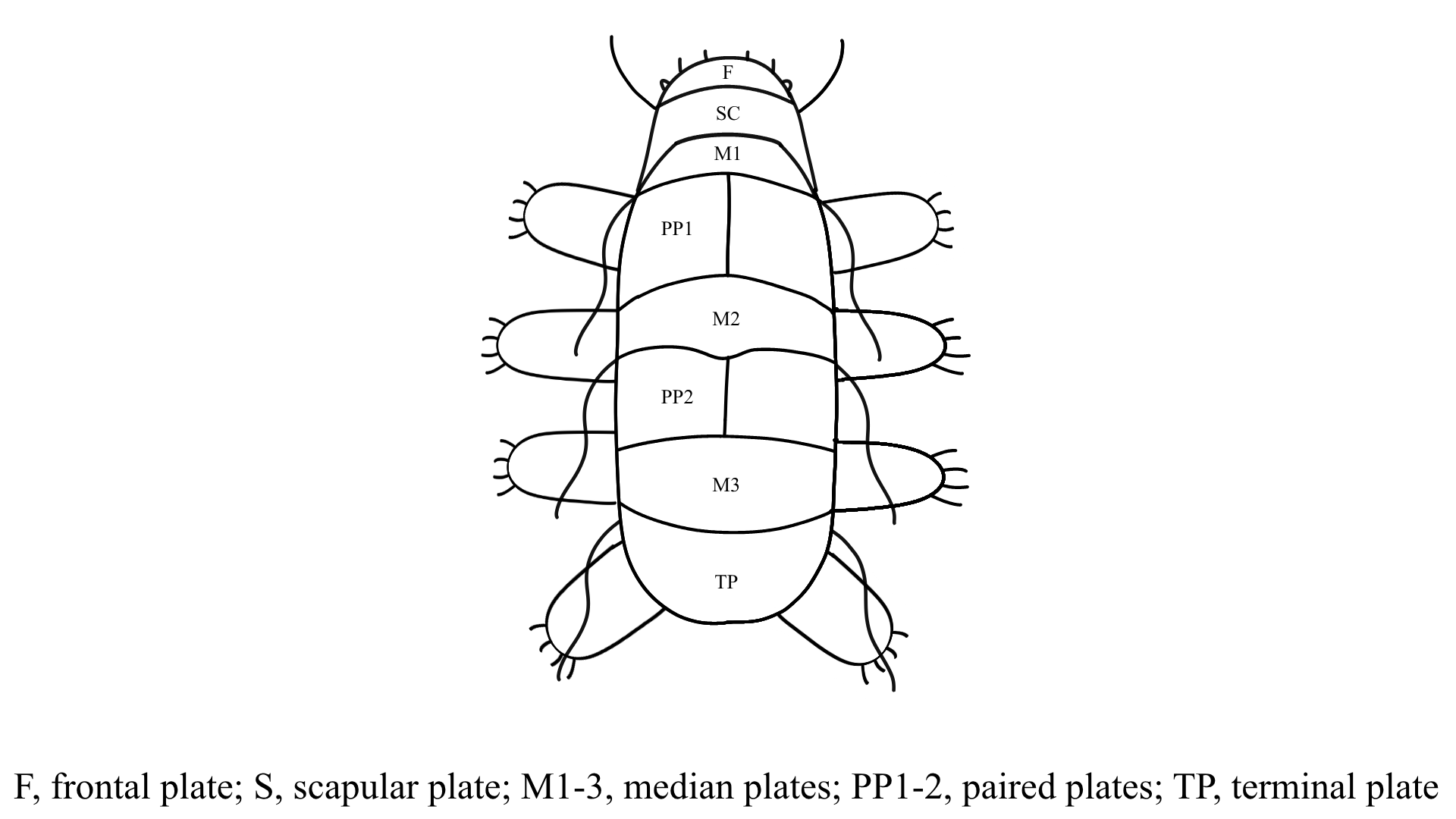
Armoured (dorsal) plates?

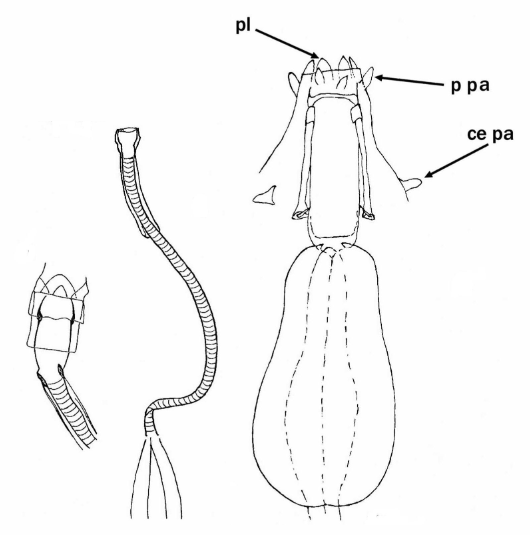
Pharyngeal tube flexible?

Note: in a tardigrade with snout extended, the flex of the pharyngeal tube may not be obvious.


Images from Pilato G, Binda MG. 2010. Definition of families, subfamilies, genera, and subgenera of the Eutardigrada, and keys to their identification. Zootaxa. 2404: 1-54.

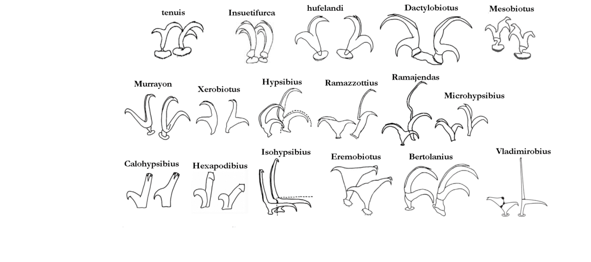
Claw type

tenuis: With stalk, a long common tract formed by the joined primary & secondary branches, and distal sections of both the secondary branch – shorter than the primary – and primary branch forming almost a right angle.
Insuetifurca: Modified hufelandi: Basal section subdivided into a very short stem & a large distal portion.
hufelandi: Primary & secondary branches rigidly joined, forming an acute angle, with variable but long common basal tract and cylindrical stalk.
Dactylobiotus: Basal section a trapezoidal lamina; secondary branch clearly shorter than primary and inserted near the base of the latter; the two branches form an almost right angle.
Mesobiotus: Primary & secondary branches rigidly joined, forming acute angle, with variable but long common basal tract with internal septum defining a distal part, with cylindrical stalk. Often very prominent, strong accessory point on primary.
Murrayon: Basal section a trapezoidal lamina; primary & secondary branches joined to each other for a very short portion and form an acute angle.
Xerobiotus: Whole double claw equally sclerified; basal section not subdivided into a basal stem & distinct distal portion; no separating septa present; primary & secondary branches rigidly joined forming an acute angle.
Hypsibius: Secondary branch forming a continuous curve with its basal tract; in external claws the primary branch connected to the basal tract with a flexible part while the inner claw is rigid.
Ramazzottius: External claws with basal section longer than the secondary branch; primary branch very long and slender and connected to basal tract with an evident, thin, flexible tract; internal claws short & stout, rigid.
Ramajendas: External claws similar to Hypsibius type with extremely long and slender primary branch; internal claw similar to Isohypsibius type.
Microhypsibius: Small, rigid, with an evident thin basal tract continuous with the primary branch; secondary branch rigidly joined to the primary branch at a distance from the base of the claw.
Calohypsibius: Small, rigid, in frontal view with a base as large as the sum of the primary and secondary branch widths, without a suture between the two branches.
Hexapodibius: Very short, without common basal tract, with base as large as the sum of the primary and secondary branches.
Isohypsibius: Secondary branch inserted perpendicularly on the claw basal tract.
Eremobiotus: Isohypsibius-type very modified with two branches of each claw joined to one another for a long portion of their length constituting a large common tract from which the two distal tracts diverge forming an angle of almost 180°.
Bertolanius: Subdivided into three distinct sections: basal section, secondary branch and primary branch (rigidly joined to the secondary branch), one on top of the other and separated by septa; the angles between the main and secondary branch different in external and internal claws – and acute angle (about 45°) is formed by the external claw, and an almost right angle (about 80°) by the internal claw; internal claws can rotate on their base up to 180°.
Vladimirobius: external at least 2x length of internal (measured from base of claw), internal very wide at the junction of primary and secondary branches, short and thin basal part, primary branches with very prominent gibbosity-like projection near the junction of primary and secondary branches; external very thin with long basal part; accessory points of both claws very near to end of primary branch.
Calcarobiotus: Basal section, with or without basal spurs, subdivided into a thin flexible stem & a wide distal section in the shape of an upside-down triangle distally delmited by a septum; primary & secondary branches similar in shape & size.
Macroversum: Basal section subdivided into a thin, flexible, stem and distal portion not very sclerotized; primary & secondary branches connected to each other for a short portion and form an almost right angle.

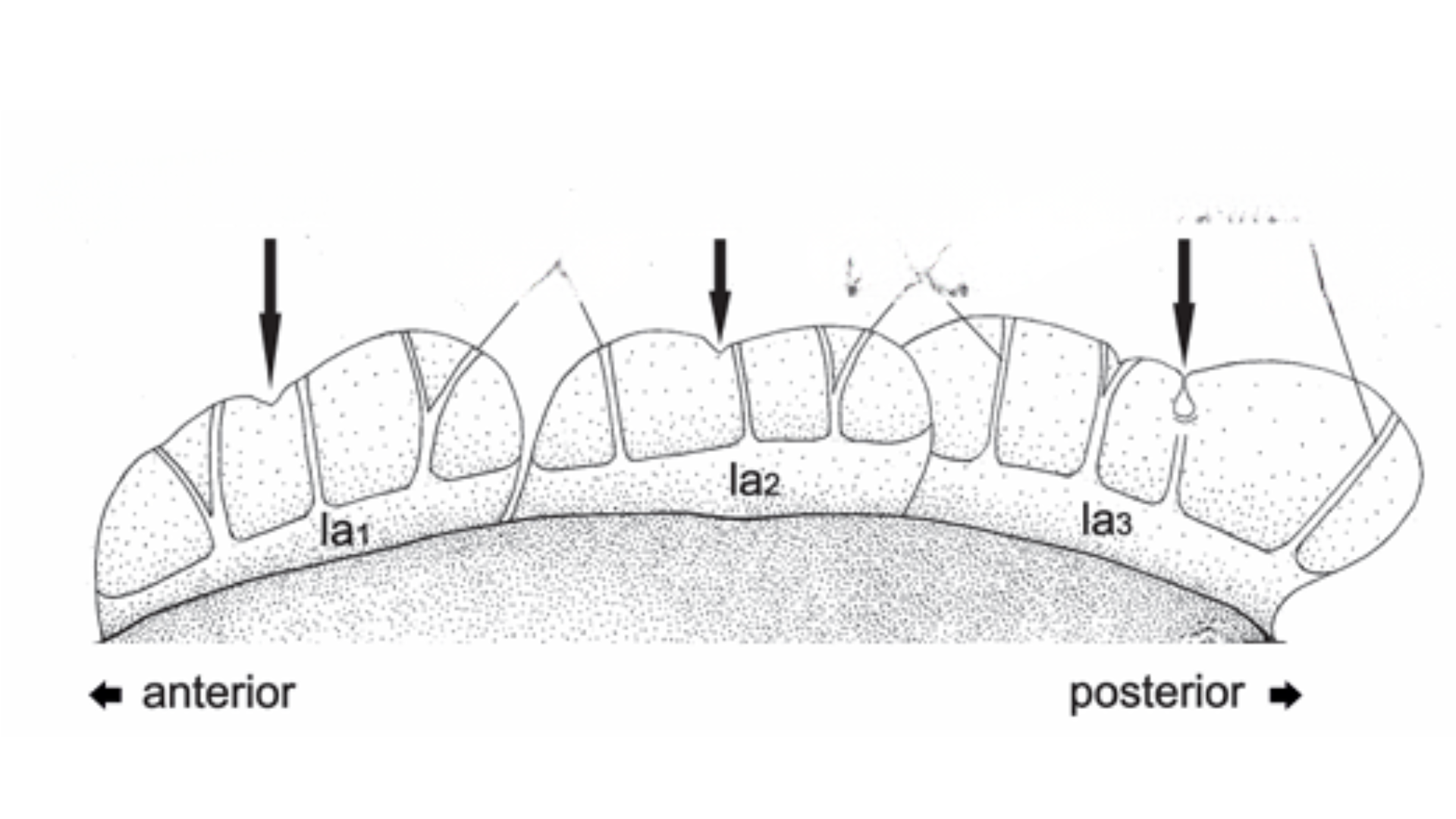
Number of lateral alae

Hansen, J.G. & Kristensen, R.M. (2021) A new genus and five new species of the subfamily Florarctinae (Tardigrada, Arthrotardigrada). European Journal of Taxonomy, 762, 149-184.

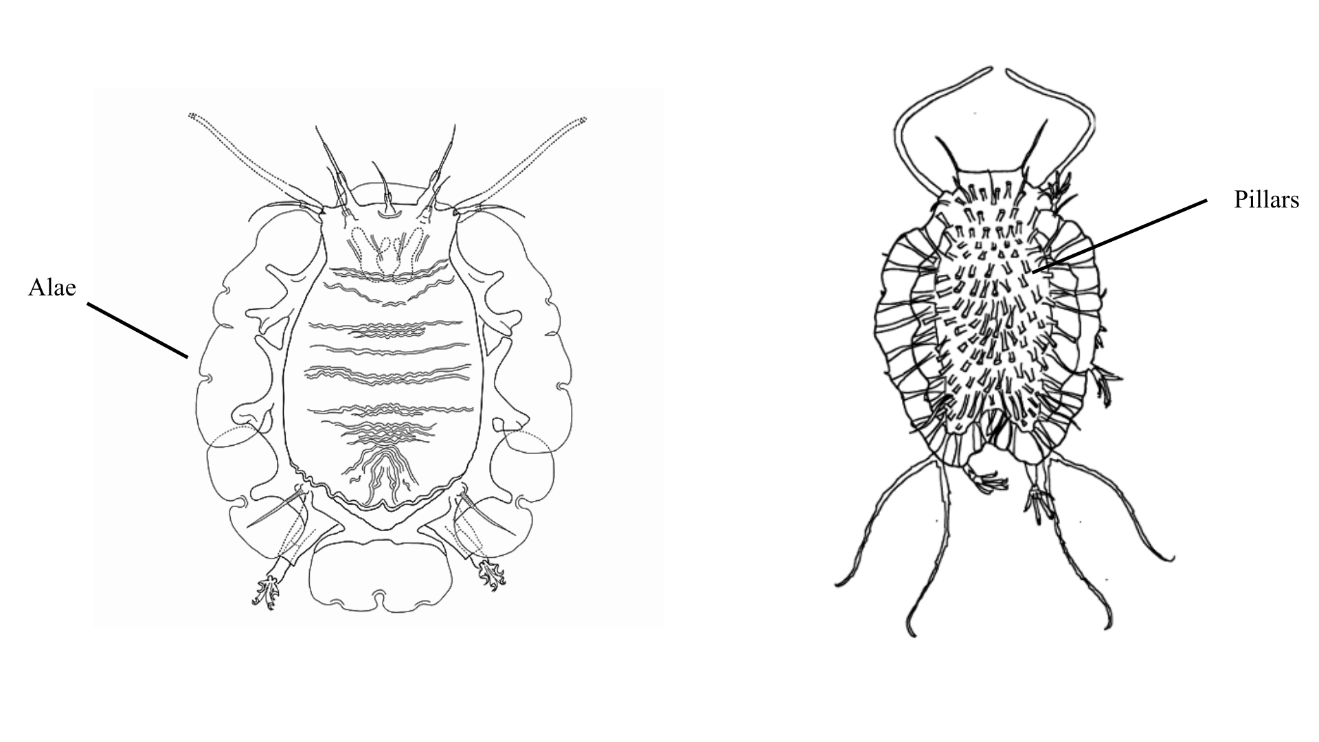
Epicuticle

Left image: Anguas-Escalante, A., De Jesús Navarrete, A., Demilio, E., Pérez-Pech, W.A. & Hansen, J.G. (2020). A new species of Tardigrada from a Caribbean reef lagoon, Florarctus yucatanensis sp. nov. (Halechiniscidae: Florarctinae). Cahiers de Biologie Marine, 62, 377-385.

Right image: Fontoura, P., Bartels, B.J., Jørgensen, A., Kristensen, R.M. & Hansen, J.G. (2017) A dichotomous key to the genera of the marine Heterotardigrades (Tardigrada). Zootaxa, 4294, 1-45.

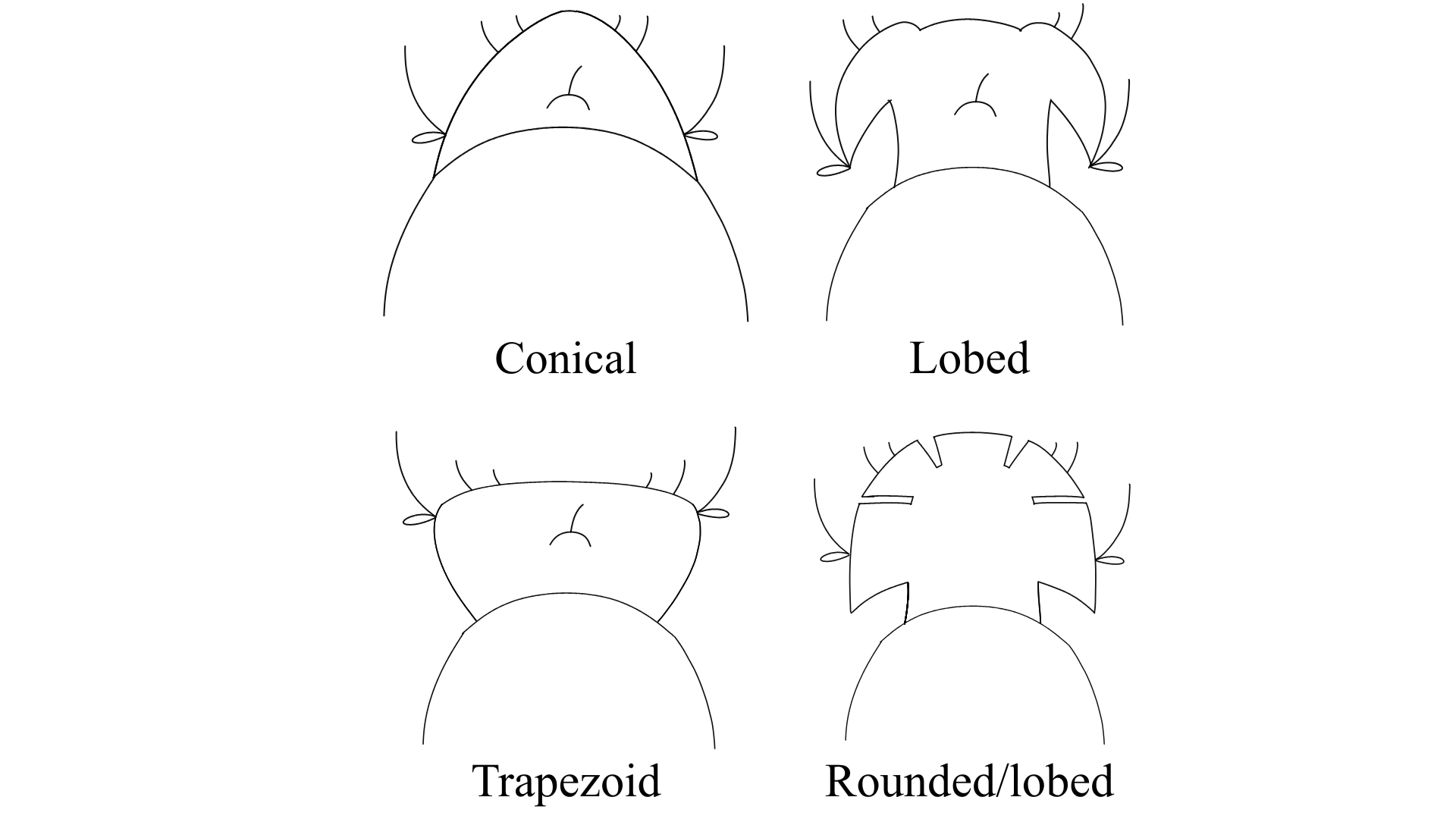
Head Structure

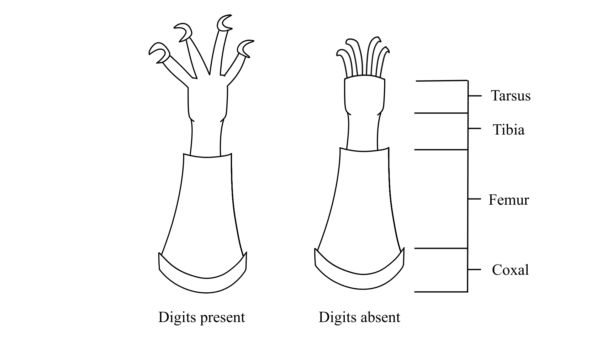
Digits

Note: If specimen has pads instead of digits, select “Pads”

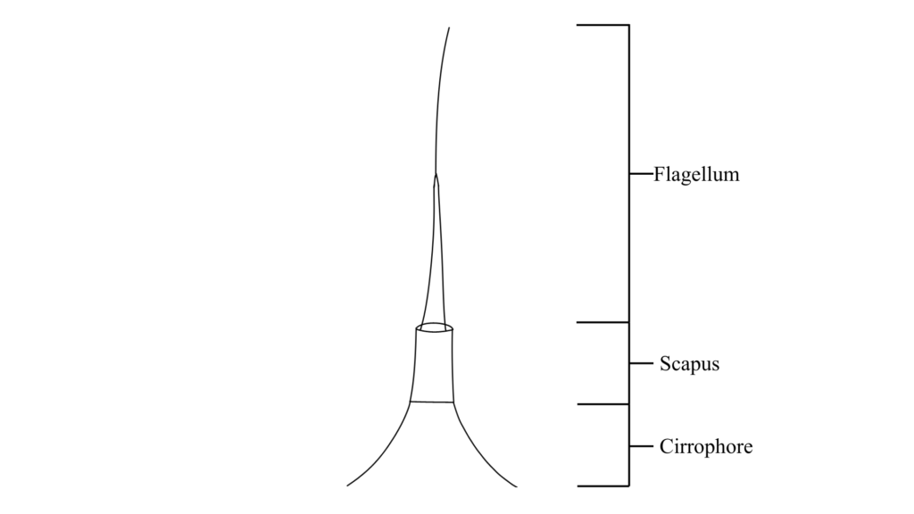
Cephalic cirri flagellum form

Modified image from Renaud-Mornant 1982 0708 Renaud-Mornant J. 1982. Species Diversity in Marine Tardigrada. Proceedings of the Third International Symposium on the Tardigrada. 149-178

Note: If unsure of form, select “Present”

Cephalic cirri scapus form

Modified image from Renaud-Mornant 1982 0708 Renaud-Mornant J. 1982. Species Diversity in Marine Tardigrada. Proceedings of the Third International Symposium on the Tardigrada. 149-178

Note: If unsure of form, select “Present” 

Cephalic cirri cirrophore form

Modified image from Renaud-Mornant 1982 0708 Renaud-Mornant J. 1982. Species Diversity in Marine Tardigrada. Proceedings of the Third International Symposium on the Tardigrada. 149-178

Note: If unsure of form, select “indistinct” for cirrophore

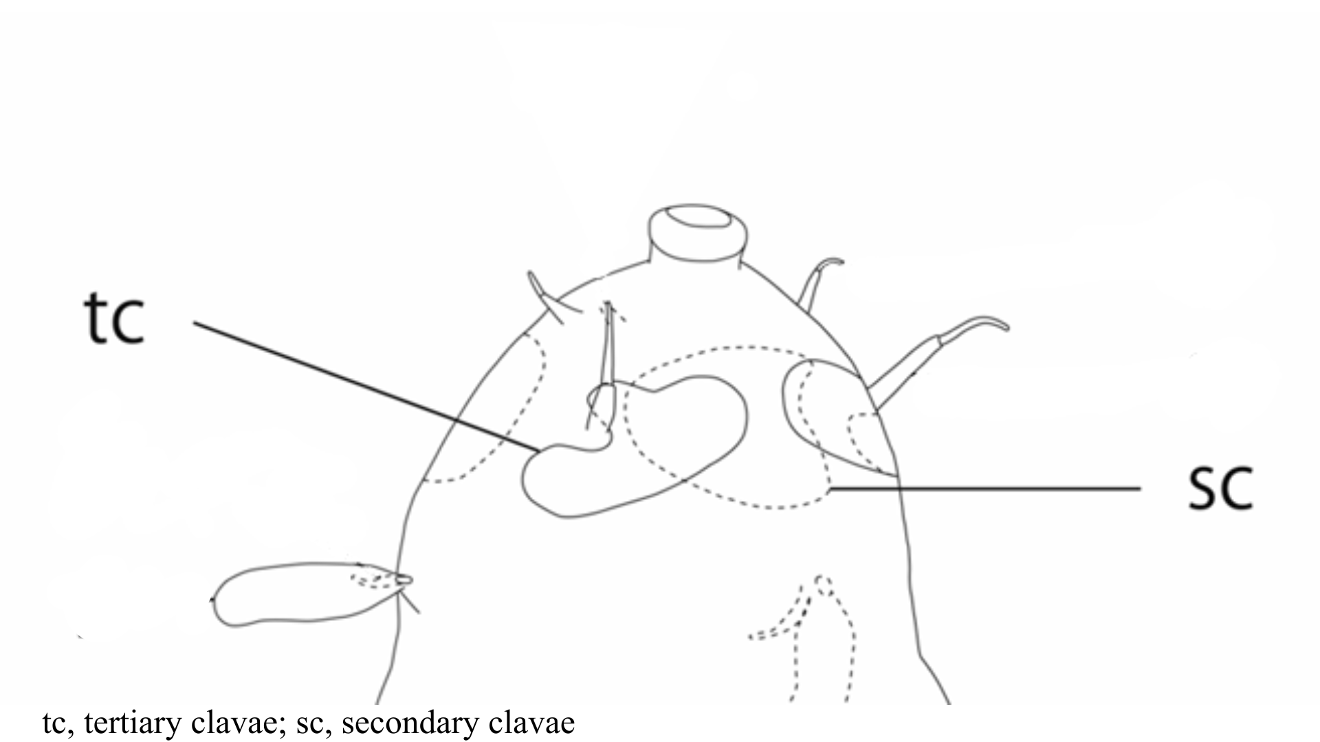
Secondary and tertiary clavae

Image from Fujimoto S, Hansen JG. 2019. Revision of Angursa (Arthrotardigrada: Styraconyxidae) with the description of a new species from Japan. European Journal of Taxonomy. 150: 1-19.

Note: Secondary clavae can have similar forms to primary clavae. However, secondary clavae can be flat structures of varying shapes around the mouth cone area

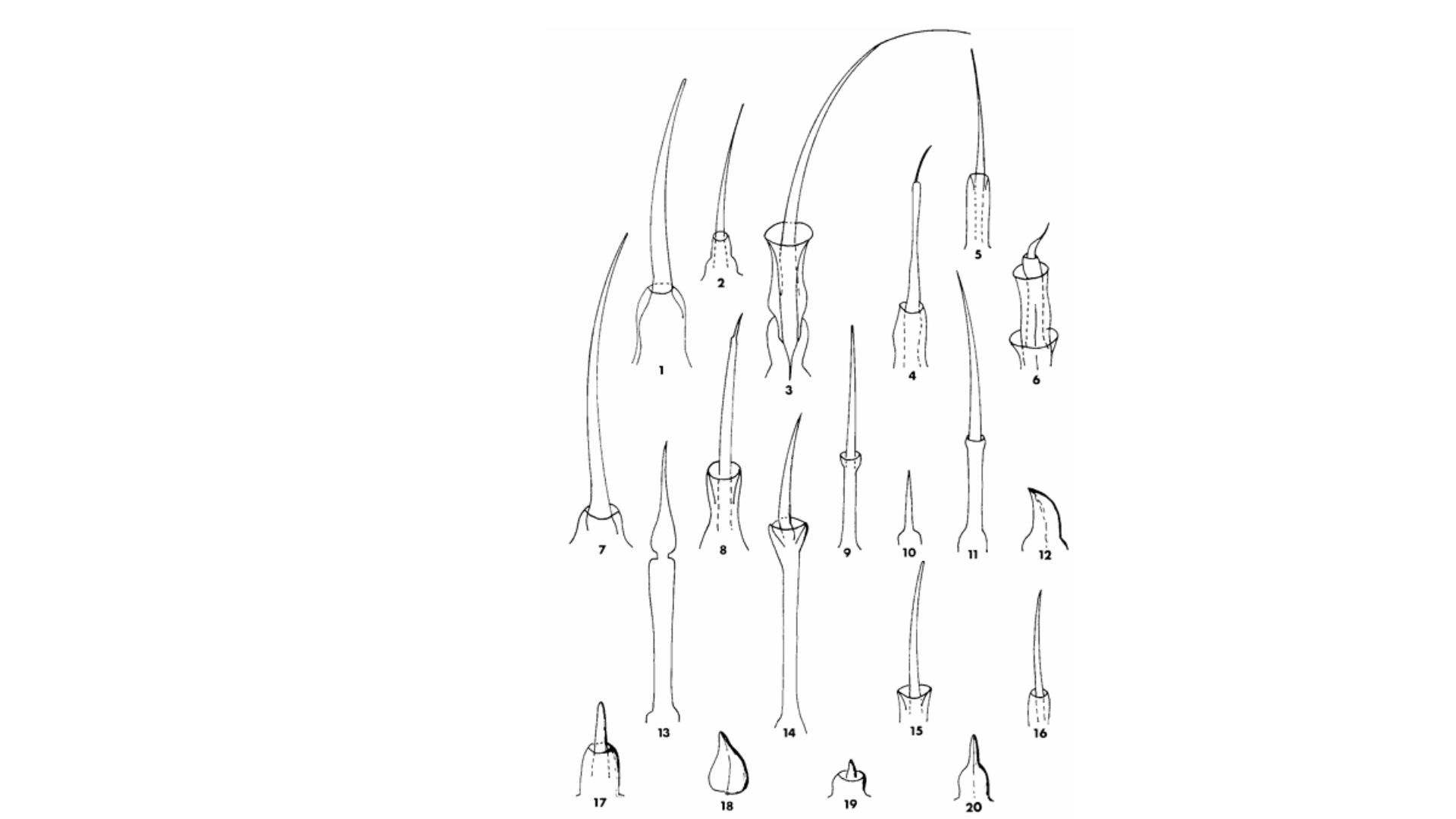
Generalized forms of primary clavae

Arthrotardigrada generalized primary clavae form

Note: Clavae forms can vary between species

Armoured plates?

Note: Select “Ventral plates” if visible.

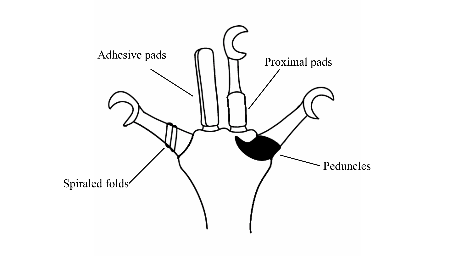
Additional digit accessory structures

Arthrotardigrada digit accessory structures

Note: Specimen can have some or none of these traits. Select those that apply.

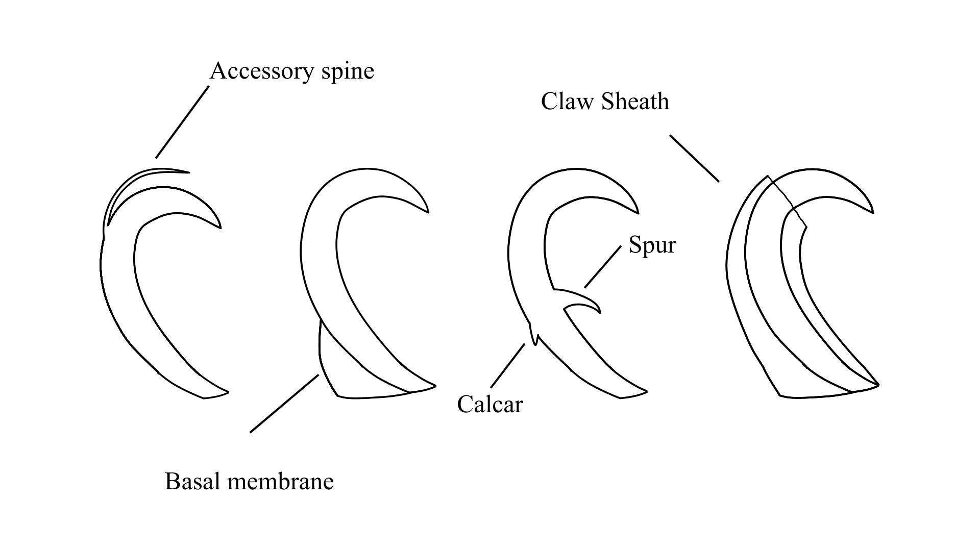
Additional claw accessory structures

Arthrotardigrada claw accessory structures

Note: Specimen can have some or none of these traits. Select those that apply.

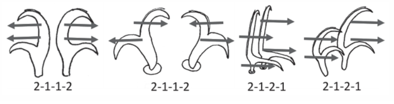
Claw Symmetry Relative To Median Plane Of Leg?

Image from Bingemer J, Hohberg K. 2017. An illustrated identification key to the eutardigrade species (Tardigrada, Eutardigrada) presently known from European soils. Soil Organisms. 89 (3): 127-149.

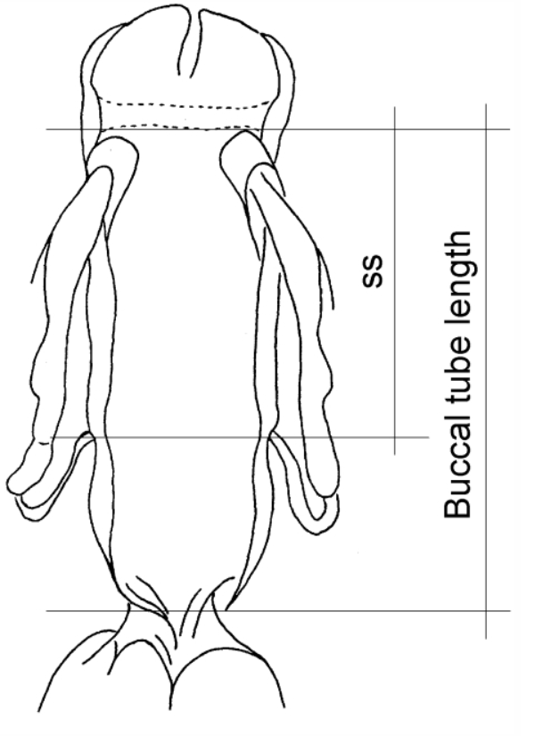
Stylet Support Insertion Point As Percentage Of Buccal Tube Length

Stylet support insertion point = ss divided by Buccal tube length, as %. Note anterior measurements begin at anterior margin of stylet sheaths, ss is centred where stylet supports reach buccal tube (this requires a good dorsal or lateral view for proper measurement)


Image from Tumanov DV. 2006. Five new species of the genus Milnesium (Tardigrada, Eutardigrada, Milnesiidae). Zootaxa. 1122: 1-23.

Buccal Tube Long, Mouth At Anterior Of A Protrusible Snout

Images from Pilato G, Binda MG. 2010. Definition of families, subfamilies, genera, and subgenera of the Eutardigrada, and keys to their identification. Zootaxa. 2404: 1-54.

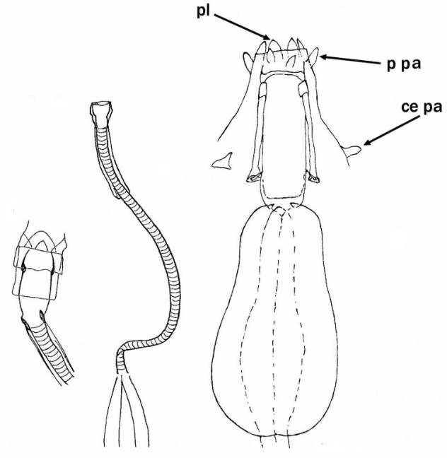
Pharyngeal Tube With Spiral Reinforcement

Images from Pilato G, Binda MG. 2010. Definition of families, subfamilies, genera, and subgenera of the Eutardigrada, and keys to their identification. Zootaxa. 2404: 1-54.

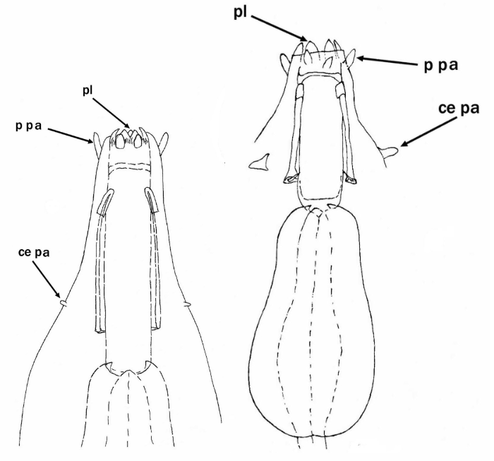
Peribuccal papillae?

Images from Pilato G, Binda MG. 2010. Definition of families, subfamilies, genera, and subgenera of the Eutardigrada, and keys to their identification. Zootaxa. 2404: 1-54.

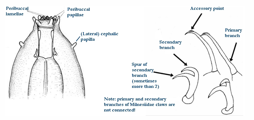
(Lateral) cephalic papillae, peribuccal papillae, Milnesiidae - type claws, exceptionally wide (and usually short) buccal tube?

Cephalic image modified from Nelson DR, Guidetti R, Rebecchi L. 2009. Tardigrada. Ch. 14 in Ecology and Classification of North American Freshwater Invertebrates, Thorp JH, Covich AP (eds.), 3rd ed.

Claw image modified from Pilato G, Binda MG. 2010. Definition of families, subfamilies, genera, and subgenera of the Eutardigrada, and keys to their identification. Zootaxa. 2404: 1-54.

Pharyngeal tube flexible?

Note: in a tardigrade with snout extended, the flex of the pharyngeal tube may not be obvious.


Images from Pilato G, Binda MG. 2010. Definition of families, subfamilies, genera, and subgenera of the Eutardigrada, and keys to their identification. Zootaxa. 2404: 1-54.

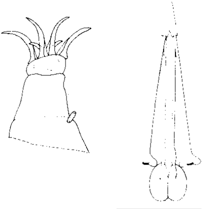
Serrated / dentate cuff / collar on legs IV

Left image from Kristensen RM. 1987. Generic revision of the Echiniscidae (Heterotardigrada), with a discussion of the origin of the family. pp. 261-335 in Bertolani R (ed). Biology of Tardigrades: Selected symposia and monographs.

Right Image from Richters F. 1926. Tardigrada. in Krumbach T. 1927. Handbuch der Zoologie, 3rd band, Walter de Gruyter & Co.

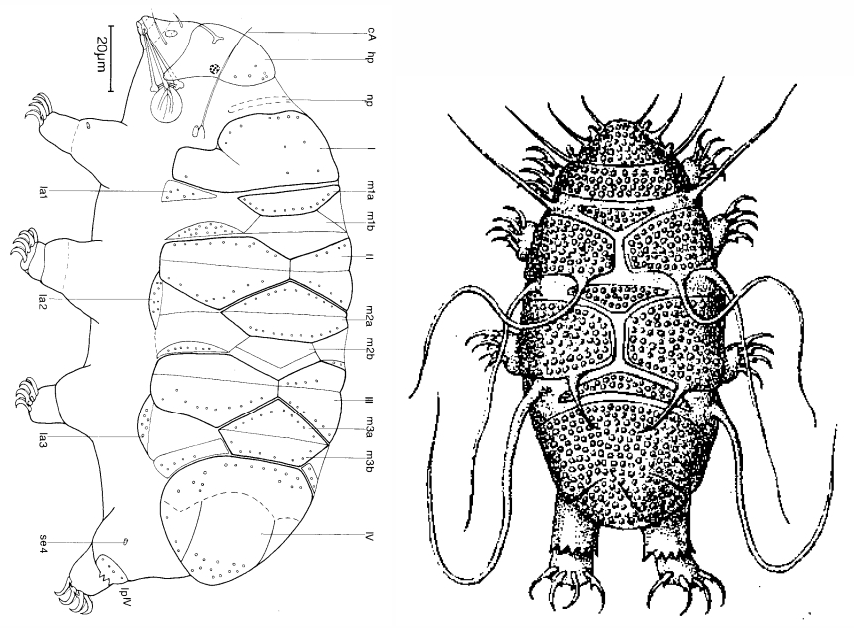
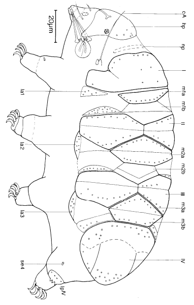
Leg sensory structures

Typically papilla on leg IV (se4 in image) and variable shape on leg I


Image from Kristensen RM. 1987. Generic revision of the Echiniscidae (Heterotardigrada), with a discussion of the origin of the family. pp. 261-335 in Bertolani R (ed). Biology of Tardigrades: Selected symposia and monographs.

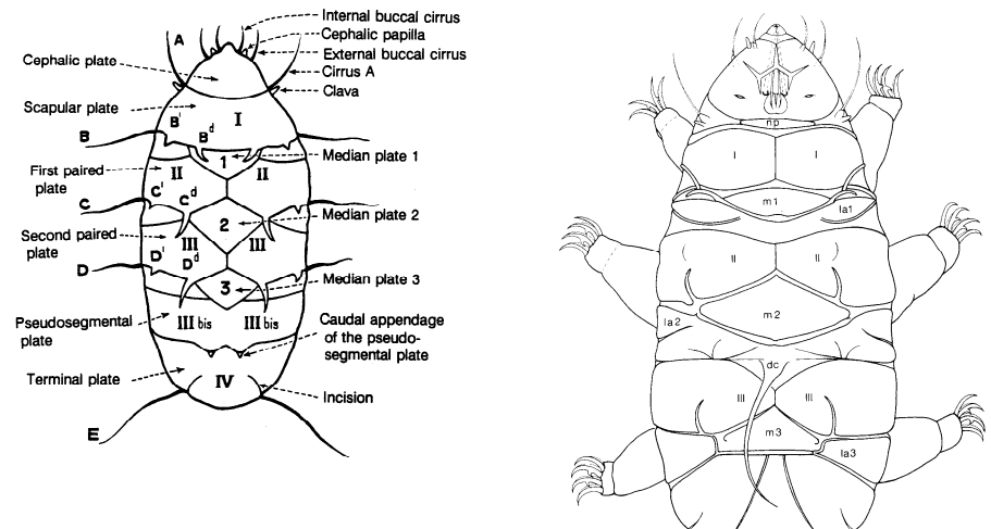
Trunk appendages

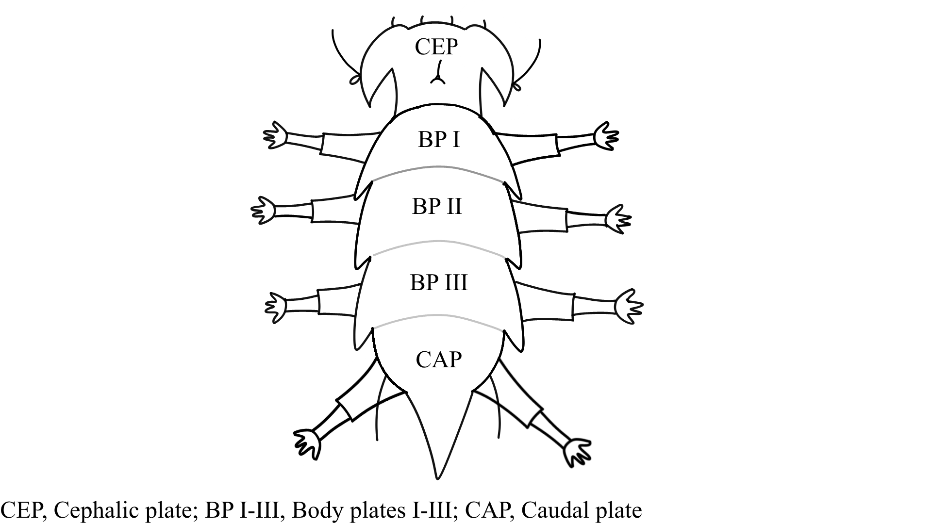
Appendages (after Cirrus A) labeled according to plate … B (Scapular plate), C (First segmental plate), D (Second segmental plate), E (terminal plate). “B” is lateral, “Bd” is dorsal

Be careful! There can be great variability within a population, with individuals (especially juveniles) lacking some appendages. When in doubt, focus on other characters first!


Left image from Ramazzotti G, Maucci W. 1983. Il phylum Tardigrada(III edizione riveduta e aggiornata). English translation by C. W. Beasley, 1995. Memorie dell’ Istituto Italiano di Idrobiologia 41: 1-1012.

Right image cropped from Kristensen RM. 1987. Generic revision of the Echiniscidae (Heterotardigrada), with a discussion of the origin of the family. pp. 261-335 in Bertolani R (ed). Biology of Tardigrades: Selected symposia and monographs.

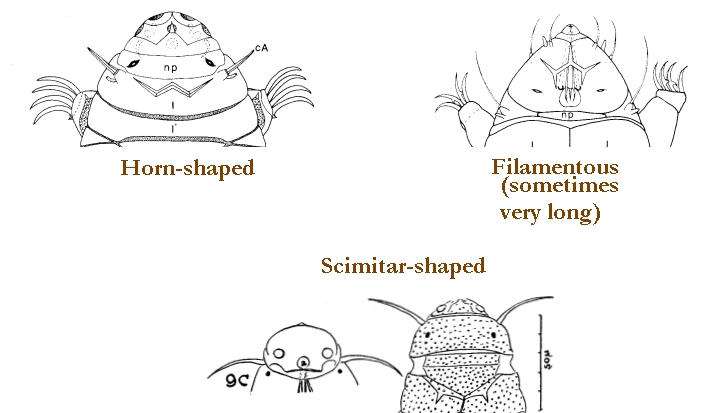
Cirrus A form

Images modified from Kristensen RM. 1987. Generic revision of the Echiniscidae (Heterotardigrada), with a discussion of the origin of the family. pp. 261-335 in Bertolani R (ed). Biology of Tardigrades: Selected symposia and monographs.

Lower image (Mopsechiniscus) modified from du Bois-Reymond Marcus E. 1944. Sobre tardigrados brasileiros. Communicaciones Zoologicas del Museo de Historia Natural de Montevideo. 1(13): 1-19 plus plates.

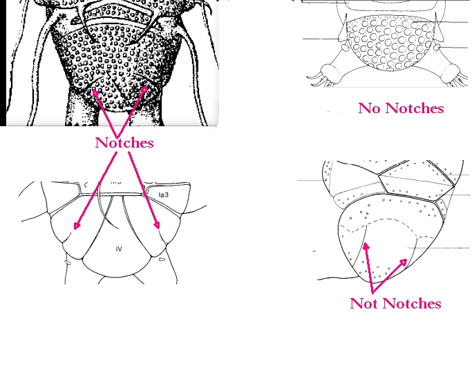
Terminal / caudal plate with two notches?

Images modified from Kristensen RM. 1987. Generic revision of the Echiniscidae (Heterotardigrada), with a discussion of the origin of the family. pp. 261-335 in Bertolani R (ed). Biology of Tardigrades: Selected symposia and monographs.

First image (Echiniscus) modified from Richters F. 1926. Tardigrada. in Krumbach T. 1927. Handbuch der Zoologie, 3rd band, Walter de Gruyter & Co.

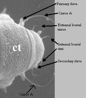
Inner & outer buccal cirri present

Image modified from Nelson DR, Guidetti R, Rebecchi L. 2009. Tardigrada. Ch. 14 in Ecology and Classification of North American Freshwater Invertebrates, Thorp JH, Covich AP (eds.), 3rd ed.

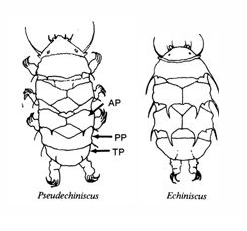
Pseudosegmental plate between last medial plate & terminal plate?

Note on 2nd image, what you see before the terminal plate is either no plate, or a full-width medial plate; don’t confuse the latter with a pseudosegmental plate!
Also, sometimes the pseudosegmental plate is paired, like segmental plates II & III. It’s still pseudosegmental!

Images from Clifford HF. 1991. Aquatic invertebrates of Alberta. University of Alberta Press, Alberta, Canada.

Segmental plates larger than intersegmental?

Image modified from Lindahl & Balser (1999), http://www.iwu.edu/~tardisdp/Keypage35.html

Any Paired Plates?

Modified from Lindahl K, Balser S.  1999. Key to tardigrade genera [Internet]. Available from: https://sun.iwu.edu/~tardisdp/Keypage33.html